FIGURE SUMMARY
Title

Phages from Ganges River curtail in vitro biofilms and planktonic growth of drug resistant Klebsiella pneumoniae in a zebrafish infection model

Authors
Sundaramoorthy, N.S., Thothathri, S., Bhaskaran, M., GaneshPrasad, A., Nagarajan, S.
Source
Full text @ AMB Express

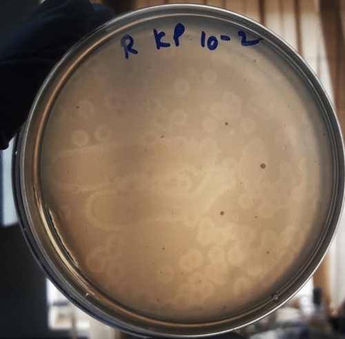
Plaque morphology of the Klebsiella pneumoniae specific phages isolated from Ganges river (KpG) The Ganges water from Rishikesh was enriched with the host and plated by agar overlay method. KpG shows depolymerase activity, which is evident from the halo around the plaques

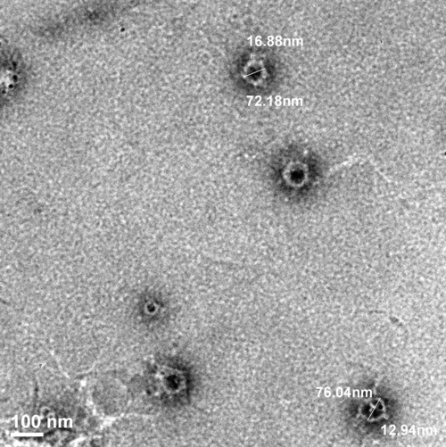
TEM analysis showed that KpG phages belonged to the family Podoviridae. Triple purified Phage lysate of high titer was stained with 2% uranyl acetate and visualized under FEI transmission electron microscope (Model JEM 2100F Jeol, Japan). Image presented is representative of multiple images

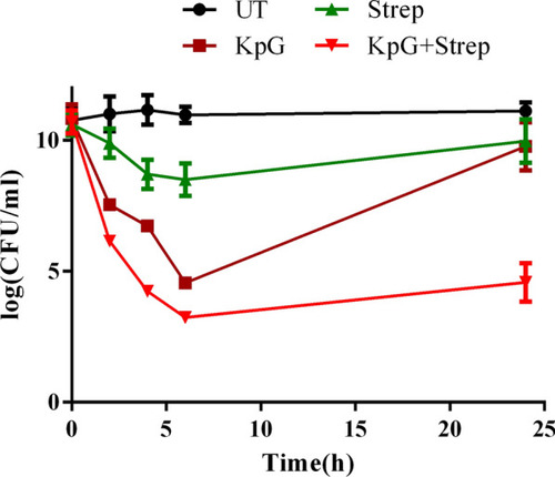
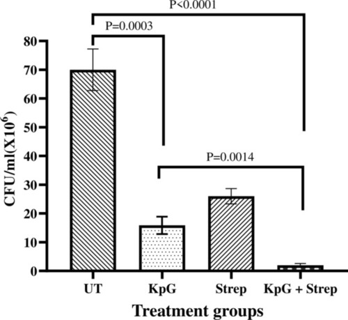
KpG phages in combination with  Streptomycin caused discernible reduction in colony counts in a time kill assay. 106 CFU/ml of K. pneumoniae was inoculated along with 108 PFU/ml of KpG phages and Streptomycin (individually and in combination). Samples were withdrawn at 0, 2, 4, 6 and 24 h serially diluted and plated on to LA plates to determine colony counts. Experiments were performed in triplicates and error bar represent standard error of the mean

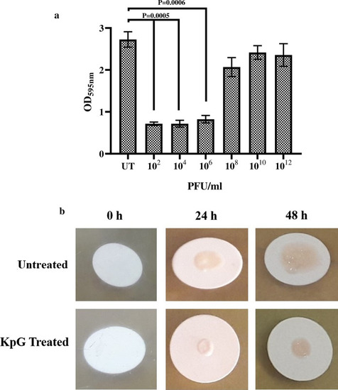
KpG phages efficiently inhibited biofilm formation of K. pneumoniae at liquid–air and at solid-air interface. a) Biofilms were formed in micro titer plates with or without KpG phages and washed with PBS. 24 h post treatment, Crystal violet was added and after 15 min, the unbound crystal violet was removed and the stain was extracted by acetic acid and the absorbance was measured at 595 nm, b) Membrane filters were placed on BHI agar and inoculated with bacteria with or without KpG. The ability of phages to decrease biofilm formation can be examined visually till 48 h. Images are representative of three independent experiments

KpG phages reduced bacterial bioburden in infected fish. 10 µl of K. pneumoniae (106 CFU/ml) were injected intramuscularly in zebrafish and 2 h post infection, KpG was administered intramuscularly. 24 h post infection, the muscle was dissected, serially diluted and plated on to LA plates. CFU/ml was calculated. Experiment was performed in triplicates and error bar represent the standard error of the mean

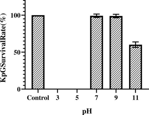
Stability of KpG at different pH. 107 PFU/ml of purified phage lysate was incubated in buffers of varying pH 3.0, 5.0, 7.0, 9.0 and 11.0 for 1 h. Phage titer was estimated using agar overlay method and survival percentage was calculated using phage titers measured in SM buffer (pH 6.8) as control. The experiment was performed in triplicates

Acknowledgments
This image is the copyrighted work of the attributed author or publisher, and ZFIN has permission only to display this image to its users. Additional permissions should be obtained from the applicable author or publisher of the image. Full text @ AMB Express