FIGURE SUMMARY
Title

Elevated preoptic brain activity in zebrafish glial glycine transporter mutants is linked to lethargy-like behaviors and delayed emergence from anesthesia

Authors
Venincasa, M.J., Randlett, O., Sumathipala, S.H., Bindernagel, R., Stark, M.J., Yan, Q., Sloan, S.A., Buglo, E., Meng, Q.C., Engert, F., Züchner, S., Kelz, M.B., Syed, S., Dallman, J.E.
Source
Full text @ Sci. Rep.

ZFIN is incorporating published figure images and captions as part of an ongoing project. Figures from some publications have not yet been curated, or are not available for display because of copyright restrictions.

PHENOTYPE:
Fish:
Observed In:
Stage: Day 5

ZFIN is incorporating published figure images and captions as part of an ongoing project. Figures from some publications have not yet been curated, or are not available for display because of copyright restrictions.

ZFIN is incorporating published figure images and captions as part of an ongoing project. Figures from some publications have not yet been curated, or are not available for display because of copyright restrictions.

Brain-wide activity in both glyt1−/− mutants and wild type siblings is significantly reduced by exposure to propofol. Four batches of sixteen to eighteen, six-day-old larvae were placed in baskets (diagramed to the left) to enable easy transfer of larvae from system water to 10 µM propofol solutions. Half contained glyt1 siblings (A) and the other half glyt1−/− mutants (B). Larvae from each treatment were fixed and stained with pERK and tERK antibodies and z-stacks of the brain were captured on a confocal microscope. Images boxed in green correspond to baseline conditions while images boxed in purple correspond to exposure to 10 µM propofol for twenty minutes. Images show standard deviation projections of pERK, tERK, pERK/tERK, and median values. To compare activity between baseline and propofol treatments, voxels with more intense pERK/tERK at a p < 0.00005 threshold under baseline conditions are shown in green while voxels with more intense pERK/tERK in propofol are shown in purple for glyt1 siblings (A) and glyt1−/− mutants in (B). It is clear that both glyt1 siblings and glyt1−/− mutants have reduced activity at anesthetic doses of propofol as expected from their similar dose/response curves. Yellow brackets, curly brackets and arrows in median images highlight staining patterns that differ significantly between glyt1−/− mutants and their siblings. Under Baseline conditions, brackets point to a region encompassing the subpallium and preoptic that has more pronounced staining in the glyt1−/− mutants while curly brackets point to a region encompassing the optic tectum, cerebellum, and hindbrain that has more intense staining in the glyt1 siblings. Under propofol, arrows point to the area postrema region of the hindbrain that has more pronounced staining in glyt1 siblings. CB cerebellum, HB hindbrain, OB olfactory bulb, OT optic tectum, P pallium, PO preoptic, SC Spinal Cord, SP Sub-pallium, VT ventral tegmentum. Scale bars = 100 µm.

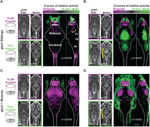
Compared to glyt1 siblings, glyt1−/− mutants show delayed activation of pre-motor, and motor regions during emergence from propofol. Four batches of sixteen to eighteen, six-day-old larvae were placed in baskets (diagramed to the left) to enable easy transfer of animals from 10 µM propofol to system water solutions. Half contained glyt1 siblings after (A) ten minutes and (B) twenty minutes in system water. The other half contained glyt1−/− mutants after (C) ten minutes and (D) twenty minutes in system water. To compare activity between propofol and wash treatments, voxels with more intense pERK/tERK at a p < 0.00005 threshold under propofol conditions are shown in purple while voxels with more intense pERK/tERK in system water during emergence are shown in green for glyt1 siblings (A, B) and glyt1−/− mutants in (C, D). glyt1 siblings show greater activation of preoptic, subpallium, and spinal cord regions under propofol, with the olfactory bulb and a regions of the diencephalon that encompasses the arousal pathways (yellow arrowhead in A) being the first brain region to become more active during emergence from propofol, followed ten minutes later by the optic tectum, cerebellum and hindbrain. glyt1−/− mutants have greater activation of preoptic and subpallium, but also optic tectum neuropil and cerebellar neuropil under propofol. As with their glyt1 siblings, the olfactory bulb is the first brain region to become activated but there is no activation of other sensory, pre-motor, and motor regions by the twenty-minute timepoint consistent with delays seen in glyt1−/− mutants recovery of locomotor behaviors after propofol. Scale bars = 100 µm.

glyt1−/− mutants’ sensory ganglia are more active while arousal and motor circuits are less active than their siblings during recovery from propofol. (A) Brain regions of interest (ROIs) that are significantly different between glyt1−/− mutants and their siblings are shown on reference brains for Baseline , Propofol, 10 min. wash and 20 min. wash conditions with sample size indicated at the base of each image. ROIs are labeled with the corresponding anatomical brain regions; an abbreviation key can be found at the end of the figure legend. (BD) Violin plots of pERK/tERK ratios for twelve different brain regions are shown with a dashed line indicating median pERK/tERK levels in baseline glyt1 siblings. For each brain region, we conducted a 2-way ANOVA for genotype and condition followed by Sidak’s multiple comparisons of genotype across conditions. Asterisks indicate p-value: *p < 0.05, **p < 0.001, ***p < 0.001, ****p < 0.0001. (A) Three sensory ganglia are shown that are elevated in glyt1 mutants during recovery. (B) Olfactory bulb and arousal pathways are shown. (C) Premotor and motor regions elevated in baseline glyt1 siblings are shown. (D) Hypnotic regions elevated in baseline glyt1−/− mutants are shown. Anatomical abbreviations: OE olfactory epithelium, subpallium SP, pallium P, Preoptic nucleus PO, Hypothalamic nucleus enriched in Qrfp-expressing neuronal cell bodies Qrfp, Ventral Thalamus VT, Dopaminergic Cluster 2 in the posterior tuberculum Dop 2, Tectum Stratum Periventriculare TSPv, Tectum Neuropil TN, Cerebellum CB, vagal motor nucleus X, posterior Lateral Line Neuromasts along the body LLN D. Scale bar = 100 µm.

ZFIN is incorporating published figure images and captions as part of an ongoing project. Figures from some publications have not yet been curated, or are not available for display because of copyright restrictions.

Acknowledgments
This image is the copyrighted work of the attributed author or publisher, and ZFIN has permission only to display this image to its users. Additional permissions should be obtained from the applicable author or publisher of the image. Full text @ Sci. Rep.