
- Title
-
TBK1-Mediated DRP1 Targeting Confers Nucleic Acid Sensing to Reprogram Mitochondrial Dynamics and Physiology
- Authors
- Chen, S., Liu, S., Wang, J., Wu, Q., Wang, A., Guan, H., Zhang, Q., Zhang, D., Wang, X., Song, H., Qin, J., Zou, J., Jiang, Z., Ouyang, S., Feng, X.H., Liang, T., Xu, P.
- Source
- Full text @ Mol. Cell
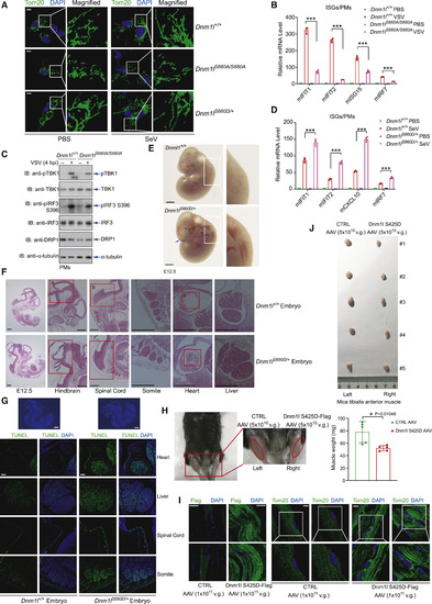
DRP1-KI Mice Reveal the Physiological Importance of the TBK1-DRP1 Axis (A) PMs isolated from DRP1-knockin (DRP1-KI) homozygotes (Dnm1l S660A) exhibited somewhat fragmented mitochondria, while PMs from Dnm1lS660D/+ chimeric mice had massively fused and elongated mitochondria under confocal microscopy. SeV-induced mitochondrial fusion was partially impeded in Dnm1l S660A PMs. Scale bar, 5 ?m. (B and C) Antiviral responses to VSV infection in Dnm1l S660A PMs was compromised, with significantly lower levels of ISG mRNAs (B) and reduced activation of TBK1 and IRF3 (C). (D) SeV-induced ISG mRNA expression in Dnm1lS660D/+ PMs was enhanced. (E) A Dnm1lS660D/+ embryo at E12.5 was largely normal, except for several hemorrhagic areas and edema in the spinal cord. Scale bars, 500 ?m. (F and G) Hematoxylin and eosin staining of Dnm1lS660D/+ embryo sections showed abnormities in spinal cord, liver, and cardiac chamber (F); TUNEL and DAPI staining revealed higher levels of apoptotic cells in organs (G). Scale bars represent 500 ?m (F and top panels of G) or 100 ?m (bottom panels of G). (H) Mouse tibialis anterior (TA) muscle had visible skeletal muscle atrophy upon AAV-9-mediated Dnm1l S425D expression at 12 weeks post-injection. (I) Dnm1l S425D protein in TA muscle was confirmed (left panels), accompanied by hyperfused and elongated mitochondria (right panels). Scale bars represent 50 ?m (left panels) or 10 ?m (right panels). (J) The volume and weight of TA muscles indicated progressive skeletal muscle atrophy in the presence of Dnm1l S425D expression. See also Figure S7. |