
- Title
-
Dare to Compare. Development of Atherosclerotic Lesions in Human, Mouse, and Zebrafish
- Authors
- Vedder, V.L., Aherrahrou, Z., Erdmann, J.
- Source
- Full text @ Front Cardiovasc Med
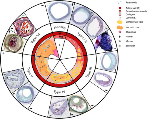
Morphology of the six types of atherosclerosis in human, mouse, and zebrafish: a schematic overview with a comparison of micrographs. Healthy arteries have an adventitia surrounding the media (M), a very thin intima (I) layer, and a large lumen (L). As atherosclerosis initiates and progresses, different types are classified as follows: Type I, intima thickening; type II, fatty streaks, with or without macrophages; type III, intermediate lesion; type IV, advanced atheroma; type V, fibroatheroma; type VI, complicated plaques with surface defects, leading to plaque rupture. The type VI mouse lesion image shows an unstable plaque in the right common carotid artery in the tandem stenosis mouse model, and it is used to represent future mouse plaque rupture models. Micrographs representing human atherosclerosis, are from Yahagi et al. ( |