
- Title
-
Low-Dose Exposure of Silica Nanoparticles Induces Neurotoxicity via Neuroactive Ligand-Receptor Interaction Signaling Pathway in Zebrafish Embryos
- Authors
- Wei, J., Liu, J., Liang, S., Sun, M., Duan, J.
- Source
- Full text @ Int. J. Nanomedicine
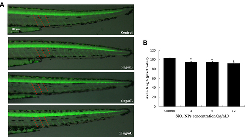
SiO2 NPs induced neurotoxicity in zebrafish. (A) SiO2 NPs disrupted the axonal integrity via the alteration in axon length in Tg (NBT:EGFP) lines. (B) Changes induced by SiO2 NPs in the axon length of zebrafish embryos were detected by NIS-Elements D 3.10 software. Data are expressed as means S.D. from three independent experiments (*p<0.05). |
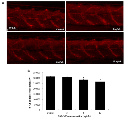
SiO2 NPs induced neurotoxicity in zebrafish. (A) SiO2 NPs caused disruption of axon pattern. (B) Changes induced by SiO2 NPs in the axon structure of zebrafish embryos were detected by NIS-Elements D 3.10 software. Data are expressed as means S.D. from three independent experiments (*p<0.05). |

The apoptosis of brain cells induced by SiO2 NPs exposure was determined using acridine orange staining in embryos of zebrafish after a 24 h exposure. (A) The images of apoptosis of brain cells were detected by fluorescence microscope. (B) The relative fluorescence of cellular apoptosis of brain was detected. Data are expressed as means S.D. from three independent experiments. |
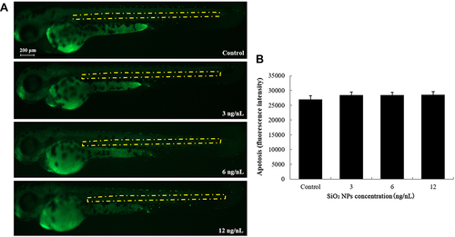
The apoptosis of central cells induced by SiO2 NPs exposure was determined using acridine orange staining in embryos of zebrafish after a 24 h exposure. (A) Central cell apoptosis images were detected by fluorescence microscope. (B) The relative fluorescence of apoptosis of central cells was detected. Data are expressed as means S.D. from three independent experiments. |