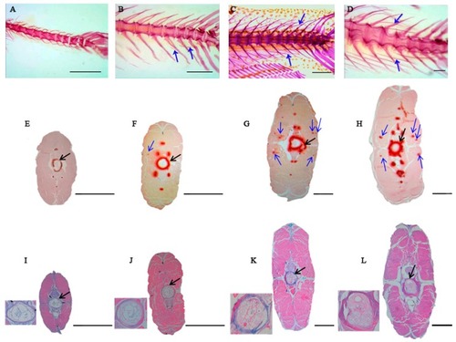
Developmental characteristics and visualization of cross sections from four key Intermuscular bone (IB) developmental stages in M. amblycephala. (AD) represent ossification process of IBs stained with ARS from S1, S2, S3, and S4, respectively. (A) S1. Body Length ≈ 11 mm (17 dph), the IBs have not emerged in the tail part; (B) S2. Body Length ≈ 16mm (24 dph), several small IBs have ossified in the tail part; (C) S3. Body Length ≈ 21 mm (29 dph), more IBs gradually ossified in the tail part with immature form; (D) S4. Body Length ≈ 32 mm (42 dph), all the IBs ossified in the tail part with mature morphology. Blue arrows indicate IBs; scale bars = 200 μm. (EH) represent histological structures of the tail part with alizarin red S staining from S1, S2, S3, and S4, respectively. (IL) represent histological structures of the tail part with alcian blue staining from S1, S2, S3, and S4, respectively. Blue arrows indicate IBs and black arrows indicate notochord sheath. The notochord sheath and vertebra were magnified in the lower left corner in (IL). Scale bars = 100 μm.

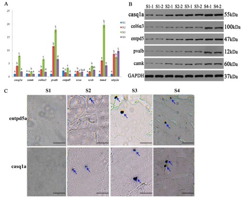
Verification of gene expression. (
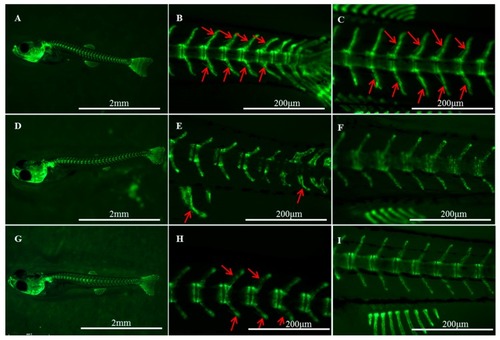
A) The relative expression of genes at different stages with qRT-PCR; for each gene, different letters above the bar of each stage indicated significant differences at the level of p < 0.05 among stages. (B) The expression levels of five proteins based on Western blot analysis; GAPDH is used as a loading control. (C) In situ expression analysis of entpd5a and casq1a genes at individual’s tail part from four stages. Blue arrows indicate IBs. Scale bar = 50 μm.

Effects of IWP-L6 (DF) and K02288 (GI) inhibitors on IB development in zebrafish larvae (Tg(sp7/GFP)). Control larvae (AC) were treated with dimethyl sulfoxide (DMSO). Red arrows indicate IBs. (B,E,H) show the tail part; (C,F,I) show the dorsal part.

Acknowledgments
This image is the copyrighted work of the attributed author or publisher, and ZFIN has permission only to display this image to its users. Additional permissions should be obtained from the applicable author or publisher of the image. Full text @ Int. J. Mol. Sci.