
- Title
-
Taking a closer look at whole organisms
- Authors
- Ichino, N., Ekker, S.C.
- Source
- Full text @ Elife
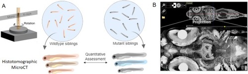
X-ray histotomography and phenotypic assessment in zebrafish.(A) At its most basic level, X-ray histotomography works by illuminating a fixed sample with a monochromatic beam of X-rays, and collecting the X-rays scattered by the sample as it is rotated. To achieve high resolution of entire organisms, Ding et al. use X-rays from a synchrotron radiation source (not shown); the scattered X-rays are converted into visible light by a scintillator and detected by a CCD camera (not shown). The combination of resolution and field of view offered by X-ray histotomography makes it possible to accurately characterize individual variations in both wild-type and mutant zebrafish at the subcellular level. (B) High-resolution image of a juvenile zebrafish (top), and an expanded view (bottom) showing details of the neural structure including individual axonal projections. This image is Figure 5—figure supplement 1 from Ding et al. |