FIGURE SUMMARY
Title

The ubiquitin ligase PHR promotes directional regrowth of spinal zebrafish axons

Authors
Bremer, J., Marsden, K.C., Miller, A., Granato, M.
Source
Full text @ Commun Biol

Robust and directional regrowth of Mauthner axons following laser-mediated transection. a, b 6-day-old double transgenic zebrafish larvae with labeling of the Mauthner neuron: Tg(hspGFF62a) and Tg(UAS:gap431-20-citrine), driving expression of membrane-bound citrine in the Mauthner using the Gal4-UAS system; bright field in (a), citrine labeled Mauthner axonal membrane along the ventral spinal cord in (b). Yellow arrowheads mark the future axon transection site at the ninth body segment. c, d In 5-day-old larval zebrafish, the Mauthner axon, labeled by Tg(hspGFF62a) and Tg(UAS:gap431–20-RFP) is surrounded by GFAP-positive glial cells in TgBAC(GFAP:GFAP-GFP) (c) and myelinating oligodendrocytes in Tg(MBP:EGFP-CAAX) (d). ek Laser-mediated transection of the Mauthner axon, intact axon before transection with the future transection site marked by a yellow arrowhead (e); retracted and sealed proximal and distal axon stump at 1 hpt (f), both at the ninth body segment. The axon stump distal to the transection site (here at the level of the anus) remains intact for several hours after transection (g) and then undergoes Wallerian degeneration, leaving behind the fragmented axon (h). Of eight axons, one fragmented earlier than 13.5 hpt and another later than 36 hpt. The mean time of fragmentation at the level of the anus for the remaining six axons was 28.1 ± 7.6 hpt. At 15 hpt, the distal stump was still intact in this example (white asterisk) and the proximal stump had started to regrow (i). Initially, the Mauthner axon regrew multidirectionally in seven of eight larvae, with some sprouts pointing caudally across the transection site; others pointed rostrally toward the cell body in the brain stem (white arrowheads; i). j, k At 48 hpt, the regrowing proximal axon had successfully crossed the transection site (yellow arrowhead). Misdirected axonal projections at the transection site were corrected (j) and the axon had regrown caudally. The entire length of the regrown axon is shown in (k). Scale bars: 200 µm in (a); 30 µm in (c), 30 µm in (e), and 30 µm in (k)

Celsr3, dync1h1, cyfip2, and phr are required for the extent of caudally directed Mauthner axonal regrowth. ah Mauthner axons were labeled in double transgenic zebrafish larvae: Tg(hspGFF62a), expressing Gal4 in the Mauthner combined with Tg(UAS:gap431–20-citrine), driving expression of membrane-attached citrine. Extent of caudally directed Mauthner axonal regrowth at 48 hpt is shown. A stitched low magnification image of the entire length of a regrown wild-type axon is shown in (a). b is the zoomed-in image of the boxed area in (a), showing the regrown wild-type axon around and caudal to the transection site. At the same magnification as the regrown wild-type axon in (b), images of celsr3, dync1h1, and cyfip2-mutant axons at 48 hpt are shown (ce), quantified in (h), demonstrating that celsr3, dync1h1, and cyfip2 are required for the extent of Mauthner axonal regrowth. At the same magnification as the regrown wild-type axon in (a), an image of a regrown phr mutant axon around and caudal to the transection site is shown in (f) and a zoomed-in image of the boxed area in (f) is shown in (g), quantified in (h), demonstrating that PHR is also required for the full extent of caudally directed Mauthner axonal regrowth. An extraspinally regrown axon branch is marked by a white asterisk (f). In addition to the caudally regrown axon, several branches have regrown rostrally (white arrows), towards the neuronal cell body in the brain stem. Transection sites are marked by a yellow arrowhead. All scale bars are 30 µm. P-values in (h) were determined using two-tailed Student’s t-test (celsr3, phr) or Mann–Whitney test (dync1h1, cyfip2). N = 21 nonmutant celsr3 siblings, n = 16 celsr3 mutants, n = 6 nonmutant dync1h1 siblings, n = 5 dync1h1 mutants, n = 8 nonmutant cyfip2 siblings, n = 8 cyfip2 mutants, n = 48 nonmutant phr siblings, n = 36 phr mutants were analyzed

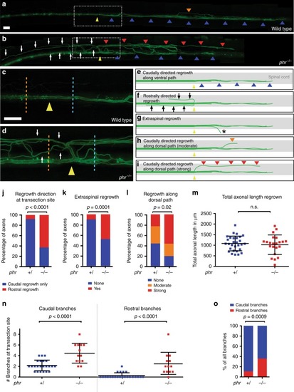
PHR promotes directional regrowth of Mauthner axons. ad Mauthner axon at 48 hpt; stitched, low magnification images of a wild type (a), and a phr mutant axon (b). Higher magnification of boxed areas (a, b) shown in c (wild type) and d (phr mutant). Yellow arrowheads mark the transection sites; white arrows mark rostrally directed projections in phr mutant. Caudally directed regrowth either labeled by blue arrowheads (along ventral spinal cord), orange arrowhead (moderate misdirected regrowth along dorsal spinal cord) or red arrowheads (strong misdirected regrowth along dorsal spinal cord). All Scale bars: 30 µm. ei Schematic representation of Mauthner axonal regrowth: Caudally directed regrowth along ventral spinal cord (blue arrowheads; e), rostrally misdirected regrowth (black arrows; f), extraspinal regrowth (asterisk in g; see also Fig. 2f) or caudally misdirected regrowth along dorsal spinal cord (red/orange arrowheads; h, i). Quantification of regrowth direction at the transection site (j). Axons regrown exclusively in the caudal direction (blue bar); axons showing additional or exclusive rostrally directed regrowth (red bar) in phr mutants (n = 37) and nonmutant siblings (n = 27). Quantification of extraspinal ventral regrowth (k). Quantification of caudally directed regrowth along dorsal spinal cord (l) using a three-category rubric of “no”, “moderate” or “strong” misdirected regrowth along dorsal spinal cord (see also e, h, i); p-values determined by Fisher exact tests (jl). N = 21 phr mutants and n = 30 nonmutant siblings were analyzed (k, l). Quantification of total length of regrown axons (sum of rostral and caudal regrowth; in n = 22 phr mutants and n = 29 nonmutant siblings with p-values determined using two-tailed Student’s t-test (m). Absolute number of rostral and caudal branches per axon in phr mutants (n = 13) and nonmutant siblings (n = 23), 40 µm rostral (orange dotted line; c, d), and 40 µm caudal (blue dotted line; c, d) to the transection site (n). Compared to nonnmutant siblings, caudally directed branches were increased twofold and rostrally directed branches were increased more than ninefold in phr mutants, (n); resulting in a higher percentage of rostrally directed branches in regrown phr mutant axons (o). P-values were determined using two-tailed Student’s t-test (N, caudal), Mann–Whitney test (N, rostral) or Fisher exact test (o)

Mutation in phr is causative for the regeneration defect in phr mutants. ac Mauthner axon at 48 hpt distal and proximal to the transection site in stitched, low magnification images of a wild-type phr+/+ axon (a), a heterozygous phrtn207b/+ axon (b) and a compound heterozygous phrtn207b/tp03 axon (c). Transection sites marked by a yellow arrowhead; rostrally directed projections in the phrtn207b/tp03 compound heterozygote are marked by white arrows (C). Scale bar for ac : 30 µm. Quantification of the extent of caudally directed regrowth in number of segments regrown caudally (d) and quantification of regrowth direction at the transection site (e), showing normal extent and directionality of regrowth in phr+/+ and phrtn207b/+ but reduced extent and misdirected regrowth in compound heterozygous phrtn207b/tp03. This shows that the regeneration deficits observed in phr mutants are indeed caused by a mutation in phr. N = 48 phr+/+, n = 53 heterozygous phrtn207b/+ and n = 13 compound heterozygous phrtn207b/tp03 were analyzed in (d, e). P-values were determined using two-tailed Student’s t-test (d), or Fisher exact test (e)

PHR is required to destabilize misdirected sprouts. Time-lapse imaging over 10 h of regrowing Mauthner axons in wild-type larvae and phr mutants, starting at 13 hpt. At 13, 18, and 23 hpt the regrown Mauthner axon of a wild-type larva (ac) and a phr mutant (df) is depicted. At 13 hpt the regrowing Mauthner axons sprouted multidirectionally in both, wild type (a) and phr mutant (d). We quantified the number of all sprouts pointing either correctly caudally (more than 90° from the original proximal projection, a subset is labeled by orange arrowheads in af) or incorrectly rostrally (up to 90° from the original proximal projection, a subset is labeled by blue arrowheads in af). Quantification is shown in g. The data show that initially both phr mutant and wild-type axons displayed rostrally and caudally directed sprouts. While the percentage of rostrally directed sprouts remained high over time in phr mutants (fg), the percentage decreased significantly in wild-type larvae at 23 hpt (c, g). Hence the percentage of rostrally directed sprouts was significantly higher in phr mutants at 23 hpt. This suggested that PHR directs regrowing axons, at least in part, by destabilizing misdirected sprouts. N = 10 phr mutants and n = 8 wild-type larvae were analyzed (one axon per fish). P-values were determined using two-tailed Student’s t-test, p-values ≤ 0.007 indicated significant differences after Bonferroni correction. Scale bar in (a) is 30 μm

PHR does not control the morphology of MBP- and GFAP-positive glial cells. Time-lapse imaging over 9 h of regrowing Mauthner axons labeled by Tg(hspGFF62a) and Tg(UAS:gap431-20-RFP). Myelinating oligodendrocytes are transgenically labeled in Tg(MBP:EGFP-CAAX) in (aj) and GFAP-positive glial cells are labeled in TgBAC(GFAP:GFAP-GFP) in (kn). In wild type (ae) and phr mutant (fj), laser-mediated axon transection caused minor damage (fragmentation and rounding) of myelinating oligodendrocytes (white stars) around the transection site (yellow arrowheads), compared to the pretransection images (e, j). Except minor damage, myelinating oligodendrocytes neither displayed obvious morphological changes nor interfered with the transection site or formed any obvious scar tissue in any of the larvae (n = 4 wild type and n = 4 phr mutants). We did not observe any differences in the morphology of MBP-positive glial cells between wild type and phr mutant larvae before or after transection. Note also the rostrally extending axonal processes in wild type (retracting over time; shorter in (d) than in (c), marked by white arrows), and in the phr mutant (longer in (i) than in (h), marked by white arrows). kn Laser-mediated axon transection also caused minor damage to GFAP-positive glial cells in all larvae analyzed (n = 4 wild type, n = 4 phr mutants). Except minor damage, GFAP-positive glial cells neither displayed obvious morphological changes nor interfered with the transection site or formed any obvious scar tissue in any of the larvae (n = 4 wild type and n = 4 phr mutants). We did not observe any differences in the morphology of GFAP-positive glial cells between wild type and phr mutant axons before or after transection. Scale bar in (a) is 20 μm

PHR controls growth cone size and filopodia length. ae F-actin (green) and microtubules (magenta) were simultaneously labeled to visualize the growth cone cytoskeleton of regrowing Mauthner axons, in double transgenic Tg(hspGFF62a) Tg(UAS:lifeact-GFP-v2a-EB3-RFP) nonmutant siblings (a) or phr mutants (b) with long F-actin-rich filopodia labeled by yellow arrowheads (b); debris of the distal axon stump marked by white asterisks (a). Laser-mediated axon transection were performed in 5-day-old larvae and regenerating axons were imaged between 12 and 15 hpt. Scale bar in (b) is 10 µm. Schematic drawing of the phr mutant growth cone, showing how the growth cone was defined and how filopodia length was measured (c). The beginning of the growth cone was defined as an increase in the axon diameter of 25% or more compared to the proximal axon shaft, which usually correlated with an obvious increase in F-actin labeling. Filopodia were measured by a straight line from filopodia base to the end of the visible F-actin signal. Growth cone size as the area above a defined intensity threshold is displayed in (d). Growth cones were significantly larger in regrowing phr mutant axons compared to nonmutant siblings. P-value was determined using two-tailed Student’s t-test. Filopodia length was determined and the percentage of filopodia below or ≥ 5 µm shown as bar graphs (e). Significantly more filopodia ≥5 µm were seen in regrowing phr mutant axons compared to nonmutant siblings. P-value was determined using the Fisher exact test. N = 14 growth cones in 5 non-mutant siblings and n = 11 growth cones in 5 phr mutants larvae were measured; n = 94 filopodia in 5 nonmutant siblings and n = 119 filopodia in 5 phr mutant larvae were measured. More growth cones and individual channels are shown in Supplementary Fig. 3

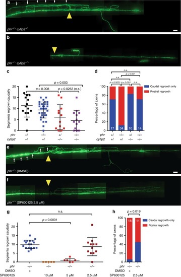
Cyfip2-deficiency and JNK inhibition rescue misdirected regrowth in phr mutant Mauthner axons. ad Laser-mediated axon transections were performed in 4-day-old larvae with different cyfip2 and phr genotypes. Regrowth was assessed 48 hpt. A stitched image of a regrown phr mutant axon is shown in (a). A yellow arrowhead marks the previous transection site. From the transection site several projections have regrown rostrally (white arrows). In contrast, in a phr−/−cyfip2−/− double mutant, there is only caudally directed regrowth (b). Scale bar: 30 µm. Quantification of extent of caudally directed regrowth (c). Quantification of percentage of axons with rostrally misdirected regrowth (d). P-value was determined using two-tailed Student’s t-test (c) or Fisher exact test (d). N of axons (1 axon per larva) for (c) and (d) as indicated in white numbers in the bar diagram in (d), except for phr+/cyfip2−/−: 12 axons were analyzed in (c) and 10 in (d), because two did not regrow at all in either direction. Only p-values ≤ 0.017 indicated significant differences after Bonferroni correction in (c). eh Laser-mediated axon transections were performed in 5-day-old phr−/ larvae. After transection, larvae were treated with DMSO (control) or with JNK inhibitor SP600125. Regrowth was assessed at 48 hpt. Stitched images of regrown phr mutant axons are shown (ef), DMSO control (e), JNK inhibitor SP600125 2.5 μM treated (f). Yellow arrowheads mark previous transection sites. From the transection site, there is a rostral trajectory (white arrows) in the DMSO-treated larva. Scale bar: 30 µm. Quantification of extent of caudally directed regrowth (g). SP600125 dose-dependently reduced Mauthner axonal regrowth. At 2.5 μM extent of Mauthner axonal regrowth was not significantly reduced. P-values were determined using two-tailed Student’s t-tests (g). Quantification of percentage of axons with rostrally misdirected regrowth in DMSO- or 2.5 μM SP600125-treated larvae (h). Number of axons (one axon per larva) in (g): DMSO: n = 18; SP600125 10 μM: n = 5; 5 μM: n = 6; 2.5 μM: n = 12. Numbers in (h): DMSO: n = 18 and SP600125 2.5 μM: n = 11 (one less than in (g) because one did not regrow at all in either direction)

Acknowledgments
This image is the copyrighted work of the attributed author or publisher, and ZFIN has permission only to display this image to its users. Additional permissions should be obtained from the applicable author or publisher of the image. Full text @ Commun Biol