
- Title
-
Methylglyoxal-Induced Retinal Angiogenesis in Zebrafish Embryo: A Potential Animal Model of Neovascular Retinopathy
- Authors
- Li, Y., Zhao, Y., Sang, S., Leung, T.
- Source
- Full text @ J Ophthalmol
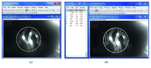
Quantification of the vascular area of retina at 4 dpf using Fiji-ImageJ software. An example of a retinal vascular image: use Fiji-ImageJ “area measurement” function under “Analyze” to measure the relative vascular vessel area over the retinal area. (a) Total retinal area by drawing a uniform circle of 120 pixel diameter = 11304 pixel area. (b) Draw and measure areas outside the vascular vessels = 2393 + 1256 + 638 + 1640 + 259 + 366 = 6552. Then, calculate % vascular vessel area of the retina = (11304 − 6552)/11304 × 100% = 42.0%. |
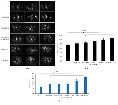
Effects of MG and glucose treatment on retinal angiogenesis in 4 dpf zebrafish. Zebrafish embryos were incubated with solution containing MG with or without glucose from 10 hpf to 4 dpf. (a) Fluorescence microscope observations of the retinal vessels of drug-treated embryos are shown at 4 dpf. The 1000 PHENOTYPE:
|

Effect of VEGF receptor inhibitor (PTK787) in MG-induced zebrafish. (a) Fluorescence microscope observations shown added 0.5 PHENOTYPE:
|