
- Title
-
Dasatinib inhibition of cSRC prevents the migration and metastasis of canine mammary cancer cells with enhanced Wnt and HER signaling
- Authors
- Timmermans-Sprang, E.P.M., Mestemaker, H.M., Steenlage, R.R., Mol, J.A.
- Source
- Full text @ Vet Comp Oncol

The effect of the rous sarcoma proto‐oncogene (cSRC) inhibitor dasatinib in zebrafish embryos. The effect of the cSRC inhibitor dasatinib on xenografted DsRed‐labelled canine mammary tumor cell line (CMT)‐U27 cells in zebrafish embryos. Three days post‐injection (3 dpi) in the yolk sac of untreated cells (A) cell proliferation in the yolk sac and cells in the tail veins were observed, whereas 10 μM dasatinib‐treated cells (B) remained in the yolk sac. At 5 dpi for the control cells (C), further yolk sac proliferation and clear extravasation in the tail veins were observed, whereas after dasatinib pretreatment, only limited survival in the yolk sac was observed (D). When the control cells were directly injected in the duct of Cuvier at 3 dpi (E) tumorous masses were observed to have formed in the tail veins, and extravasation was observed near the heart, which is an effect that was not observed in dasatinib‐treated cells (F). At 5 dpi, tumorous masses of control cells were extended (G), whereas only some of the dasatinib‐treated cells (H) ended in the tail veins but did not form tumorous masses. Statistical analysis was performed (I, J) using Fisher's exact test. *P < 0.05 was considered significant
|
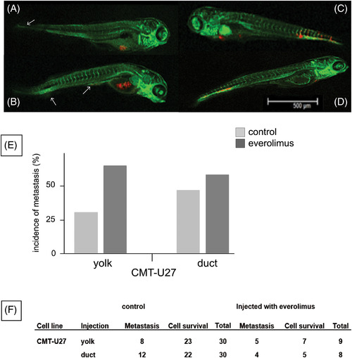
The effect of the phosphatidyl‐3‐kinase (PI3K) inhibitor everolimus in zebrafish embryos. The effect of the mammalian target of rapamycin (mTOR) inhibitor everolimus on xenografted DsRed‐labelled canine mammary tumor cell line (CMT)‐U27 cells in zebrafish embryos. Three days post‐injection (3 dpi) of untreated cells in the yolk sac (A) cell proliferation in the yolk sac and cells in the tail veins were observed, whereas after pre‐treatment with 10 μM everolimus (B), even more CMT RED cells were observed, indicating that the cells survived, proliferated and intravasated (arrows). Additionally, extravasation was more pronounced when CMT RED cells were directly injected into the circulation through the duct of Cuvier (C), showing tumour cells that formed a clump halfway through the tail, and the cells with everolimus resulted in tail vein tumours and extravasation (D). Statistical analysis was performed using (E, F) Fisher's exact test. *P < 0.05 was considered significant
|