
- Title
-
Using the Protozoan Paramecium caudatum as a Vehicle for Food-borne Infections in Zebrafish Larvae
- Authors
- Flores, E., Thompson, L., Sirisaengtaksin, N., Nguyen, A.T., Ballard, A., Krachler, A.M.
- Source
- Full text @ J. Vis. Exp.
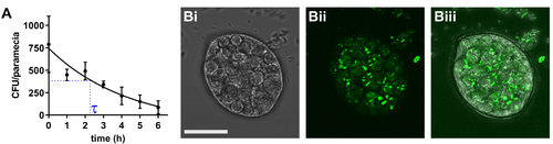
Determination of bacterial half-life in paramecia. (A) Following 2 h of co-incubation with infectious E. coli, P. caudatum was washed and transferred to medium without bacteria. At the indicated time points, numbers of viable E. coli cells were determined by dilution plating on selective agar. Results are means ± standard error of the mean (SEM; n = 3). (B) Typical image of paramecium carrying internalized bacteria, with bright field (Bi), fluorescent bacteria (Bii), and merged channels (Biii). Scale bar = 20 μm. |

Determination of zebrafish preying rate. (A) Still images from a preying video, showing a zebrafish larvae (5 dpf) preying on paramecia carrying fluorescent bacteria. Time in [seconds]. Arrow indicates the main axis of movement during striking. (B) Quantification of preying rate (paramecia intake per hour), based on n = 10 videos taken over the full 2 h exposure time. |

Colonization of zebrafish with bacteria. Zebrafish at 5 dpf were left uninfected (A) or colonized with mCherry expressing (B) E. coli or (C) S. enterica. Infection experiments may be performed in wild type (A and B) fish or transgenic lines (e.g., the line Tg(MPO::EGFP)i114 expressing green fluorescent neutrophils shown in (C). The rectal opening is marked by an arrow. (D) Higher magnification of intestinal section from whole-mount embedded larvae infected with Salmonella enterica infection. (Di) Blue = Hoechst marking nuclei, (Dii) Purple = phalloidin marking F-actin, (Diii) Red = Salmonella, (Div) merge. Scale bar = 5 μm. |