
- Title
-
V2a interneuron diversity tailors spinal circuit organization to control the vigor of locomotor movements
- Authors
- Song, J., Dahlberg, E., El Manira, A.
- Source
- Full text @ Nat. Commun.

Distribution of motoneurons and V2a interneurons. a Lateral view of the spinal cord with retrogradely labeled motoneurons by injection of Rhodamine-dextran into the muscle (left), GFP-expressing V2a interneurons (middle) and the merge of the two (right). b There were more motoneurons than V2a interneurons in each hemi-segment of the spinal cord (n = 13 animals, the boxes are bound by the 25th and 75th percentiles, whiskers extend from min. to max.). c–e Number of segmental V2a interneurons dye-coupled to slow, intermediate and fast motoneurons (n = 14 animals, error bars in the graph reflect the s.e.m.). f–h Number of intersegmental V2a interneurons in the 4 adjacent segments (2 rostral and 2 caudal) dye-coupled to slow, intermediate and fast motoneuron pools (n = 14 animals, error bars in the graph reflect the s.e.m.). Scale bars, 20 µm |

Distinct axonal projections of the two V2a interneuron types. a The bursting-type V2a interneurons had unidirectional descending axons that target the dendrites of the postsynaptic motoneurons where they produce strong excitatory drive. The filled-head black arrows indicate the sites of synaptic contacts between the V2a interneuron axon and the motoneuron dendrites. The open-head arrow reflects the fact that the axon project further than shown in this reconstruction. The dashed boxes indicate the regions that are enlarged in the lower panel showing optical sections of the sites of synaptic contacts between the V2a interneuron axon and the motoneuron dendrite that also co-localized the synaptic vesicle protein SV2 (white arrow). b The non-bursting-type V2a interneurons had bi-directional axons projecting in the caudal and rostral directions. They target the soma region of the postsynaptic motoneurons where they produce weak excitatory drive. The filled-head black arrows indicate the site of contacts between the axon of the V2a interneuron and motoneuron soma. The open-head arrows reflect the fact that the axon project further than shown in this reconstruction both in the rostral and caudal directions. The dashed box corresponds to the enlarged region in the lower panel showing an optical section where the sites of synaptic contacts between the V2a axon and the motoneuron soma as well as the localization of the synaptic vesicle protein SV2 (white arrows). c Reconstructions showing the distribution of synaptic contacts on the dendrites and soma region of the slow, intermediate and fast motoneurons from bursting-type (filled circles) and non-bursting-type (open circles) V2a interneurons. d Percentage of dendritic (filled part of the bar) versus somatic (open part of the bar) synaptic contacts onto motoneurons of the slow, intermediate and fast modules. (n = 11 slow motoneurons; n = 13 intermediate motoneurons; and n = 11 fast motoneurons). Scale bars, 10 µm, 1 µm, 20 ms, 10 mV, 0.2 mV |

Co-Localization of GFP and Chx10 in V2a interneurons.(a)Side view of the spinal cord showing that GFP-expressing V2a interneurons are Chx10-immuonoreactive. (b) Number of neurons per hemi-segment expressing GFP and Chx10-ir (n = 3 animals; error bars in thegraph reflect the s.e.m.). (c) Fraction of neurons co-localizing GFP and Chx10. Scale bar, 20 µm |
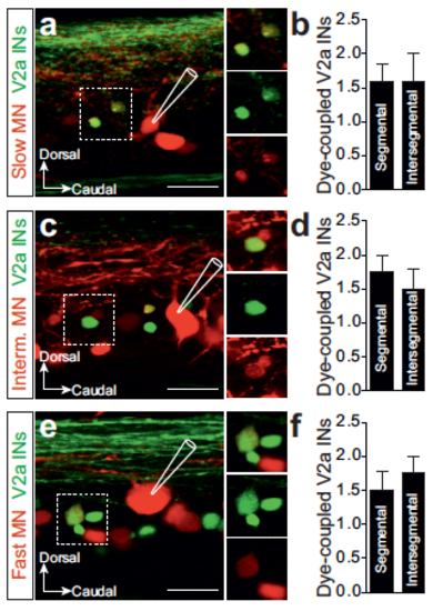
Convergence of V2a interneurons onto single motoneurons. (a) Intracellular injection of neurobiotin into a slow motoneuron resulted in dye-coupling in V2a interneurons. (b) Number of segmental and intersegmental V2a interneurons dye-coupled to single slow motoneurons (n = 5 motoneurons from separate animals, error bars reflect the s.e.m.). (c) V2a interneurons dye-coupled to an intermediate motoneuron. (d) Number of segmental and intersegmental V2a interneurons dye-coupled to single intermediate motoneurons (n=5 motoneurons from separate animals, error bars in the graph reflect the s.e.m.). (e) V2a interneurons dye-coupled to a fast motoneuron. (f) Number V2a interneurons converging onto single fast motoneurons (n=5 motoneurons from separate animals, error bars in the graph reflect the s.e.m.). Scale bars, 20 µm |