
- Title
-
Duplicated TLR5 of zebrafish functions as a heterodimeric receptor
- Authors
- Voogdt, C.G.P., Wagenaar, J.A., van Putten, J.P.M.
- Source
- Full text @ Proc. Natl. Acad. Sci. USA
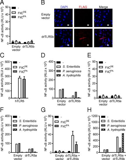
Induction of NF-κB by zebrafish TLR5b (drTLR5b) and drTLR5a upon stimulation with purified flagellins and bacterial lysates. (A and C–H) HeLa-57A cells were transfected with drTLR5b, drTLR5a, hTLR5 or drTLR5b and drTLR5a combined as indicated. Control cells were transfected with empty vector. Cells were stimulated (5 h) with vehicle (–) or 1 µg mL−1 of purified recombinant FliCSE or FliCPA flagellin (A, C, E, and G) or 2 µg mL−1 of total protein from lysates of S. Enteritidis, P. aeruginosa,or A. hydrophila (D, F, and H). Note that the NF-κB response of the drTLR5 heterodimer is higher when stimulated with bacterial lysate compared with purified recombinant flagellin. (B) Fluorescence microscopy of empty vector or FLAG-tagged drTLR5b transfected HeLa-57A cells stained with M2 α-FLAG and DAPI for nuclear visualization. (White scale bars: 10 µm.) NF-κB activity is represented by luciferase activity in RLUs. Values are the mean ± SEM of three independent experiments (A, C, E, and G) or a representative of three independent experiments (D, F, and H) all performed in duplicate. |

Effect of zebrafish UNC93B1 (drUNC93B1) on localization of drTLR5a and drTLR5b. Confocal microscopy on HeLa-57A cells transfected with (A) drTLR5-HA and drTLR5b-FLAG or (B) drTLR5a-HA and drTLR5b-FLAG and untagged drUNC93B1. Merge images show nuclei stained with DAPI (blue). White boxes in merge images indicate the magnified area shown for each channel on the right of merge images. Images were selected from three independent experiments, and three representative images are shown for each transfected group. (Scale bars in merge images: 10 µm.) |

Vesicular localization of drTLR5a and drTLR5b in the presence of drUNC93B1. Confocal microscopy on HeLa-57A cells expressing (A) drTLR5a-HA or drTLR5b-HA and drUNC93B1-FLAG or (B) drTLR5a-HA or drTLR5b-HA and untagged drUNC93B1 costained for EEA-1 or LAMP-1. Merge images show nuclei stained with DAPI (blue). White boxes in merge images indicate the magnified area shown for each channel on the right of merge images. Images were selected from three independent experiments, and three representative images are shown for each transfected group. (Scale bars in merge images: 10 μm.) |
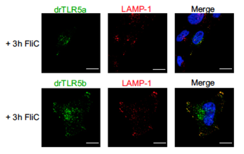
Localization of zebrafish drTLR5a and drTLR5b during flagellin stimulation. HeLa-57A cells expressing drTLR5a-HA or drTLR5b-HA and untagged drUNC93B1 were stimulated for 3 h with 1 μg mL−1 FliCSE and costained for LAMP-1. Merge images show nucleus stained with DAPI (blue). Images are representative of three independent experiments. (Scale bars: 10 μm.) |