
- Title
-
Selective IKK2 inhibitor IMD0354 disrupts NF-κB signaling to suppress corneal inflammation and angiogenesis
- Authors
- Lennikov, A., Mirabelli, P., Mukwaya, A., Schaupper, M., Thangavelu, M., Lachota, M., Ali, Z., Jensen, L., Lagali, N.
- Source
- Full text @ Angiogenesis
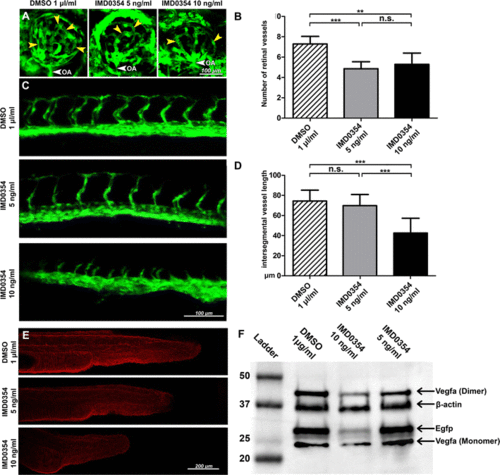
Development of retinal, intersegmental vasculature and expression of Vegf-a and EGFP in 24 h post-fertilization (hpf) Tg(fli1:EGFP)y1 zebrafish embryos treated with IMD0354. a Detection of EGFP signal from Tg(fli1:EGFP)y1 transgenic zebrafish embryos retinal vasculature at 72 hpf treated with DMSO or IMD0354 (5, 10 ng/ml). Yellow arrows indicate the retinal vessels. White arrow indicates OA (optic artery). b Quantification of the number of retinal vessels at 72 hpf with DMSO or IMD0354 treatment (5 and 10 ng/ml); (n = 7) One-way ANOVA test with Tukey multiple comparisons was used to determine statistical significance. c Detection of EGFP signal from Tg(fli1:EGFP)y1 transgenic zebrafish embryos intersegmental vasculature at 28 hpf treated with DMSO or IMD0354 (5 and 10 ng/ml). d Quantification of intersegmental vessel length (n = 16) One-way ANOVA test with Tukey multiple comparisons was used to determine statistical significance. e Immunofluorescent detection of Vegf-a (red) expression in zebrafish embryos at 24 hpf. f Western blot analysis of Vegf-a (monomeric and dimeric forms) and Egfp expression in the whole lysate of Tg(fli1:EGFP)y1 transgenic zebrafish embryos at 24 hpf, incubated with DMSO and IMD0354 (10 and 5 ng/ml). β-Actin as the loading control. n.s. p > 0.05; ***p < 0.001 |

Effect of IMD0354 treatment in zebrafish embryos and systemic effects in adult rat liver. (A) In vivo images of zebrafish embryos 24 hpf, treated with IMD0354 (0, 1, 10, 100 and 200 ng/ml). (B) In vivo images of zebrafish embryos 72 hpf, treated with IMD0354 (0, 1, 10, 100 and 200 ng/ml). (C) Mortality rate of zebrafish embryos treated with IMD0354 (0, 1, 10, 100 and 200 ng/ml) at 24 and 72 hpf. (n=40) One-way ANOVA test with Tukey multiple comparisons were used to determine statistical significance. n.s. p>0.05; *** p<0.001 (D) Hematoxylin and Eosin (H&E), (E) Cleaved-Caspase 3 (green); Nuclear counterstaining by DAPI (blue) in fluorescent images. |