
- Title
-
High-throughput imaging of zebrafish embryos using a linear-CCD-based flow imaging system
- Authors
- Liu, L., Yang, G., Liu, S., Wang, L., Yang, X., Qu, H., Liu, X., Cao, L., Pan, W., Li, H.
- Source
- Full text @ Biomed. Opt. Express
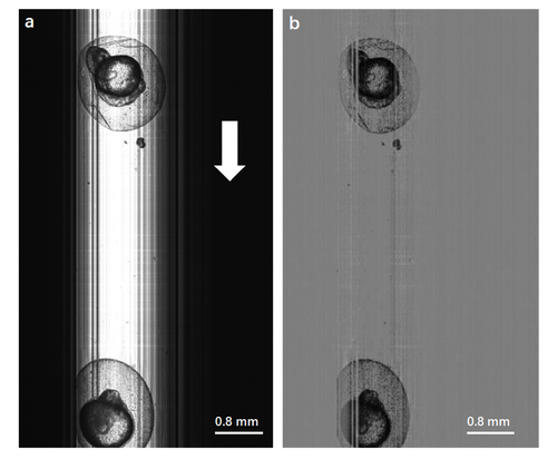
(a) Original image of zebrafish embryos obtained by Lc-FIS. The white arrow indicates the flow direction. (b) Preprocessed image after substracting the background. |
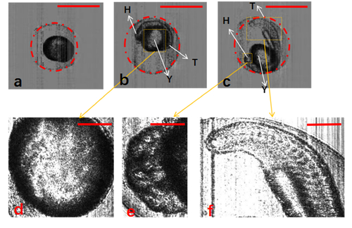
Segmentation of zebrafish embryos with chorion using Hough-transformation circle detection algorithm. (a–c) Zebrafish embryos at different development stages: 3, 13, and 21.5 hpf, respectively. (d–f) Detailed structure of yolk, head, and tail of zebrafish embryos. H, head; T, tail; Y, yolk. The scale bars in (a)–(c) represent 1 mm, and those in (d)–(f) represent 0.3 mm. |
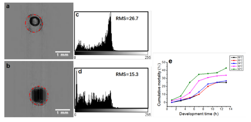
Discrimination of live and dead embryos by Lc-FIS. (a) Normal and (b) dead embryos (4 hpf) detected using the Hough-transformation algorithm. (c) and (d) Intensity distributions within the circle of the live and dead embryos, respectively. (e) Cumulative mortality rate of zebrafish embryos at temperatures of 24°C, 26°C, 28°C, 30°C, and 32°C. |
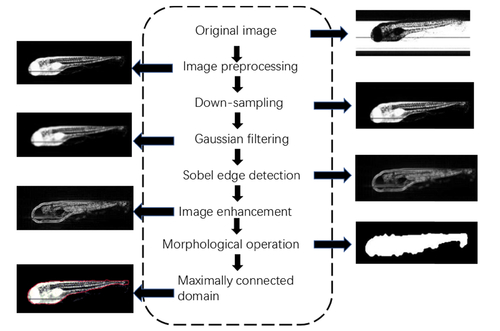
Flowchart of segmentation for zebrafish embryos without chorion. |