
- Title
-
A DNA Contact Map for the Mouse Runx1 Gene Identifies Novel Haematopoietic Enhancers
- Authors
- Marsman, J., Thomas, A., Osato, M., O'Sullivan, J.M., Horsfield, J.A.
- Source
- Full text @ Sci. Rep.
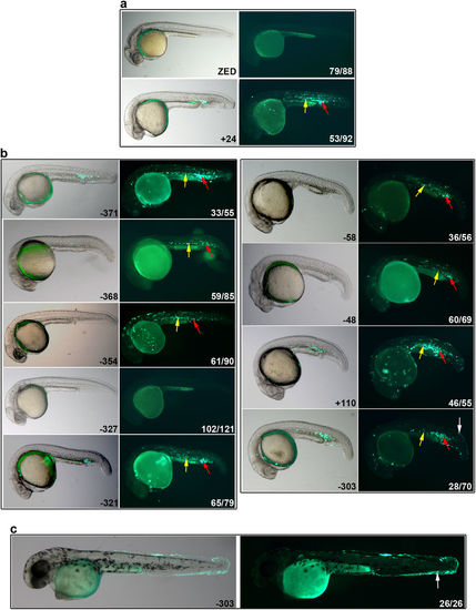
Putative mouse enhancers are active in haematopoietic regions in zebrafish. Whole mount representative lateral images of zebrafish embryos (20–24 hpf [a,b] and ~48 hpf [c]) that were injected with enhancer-GFP plasmids at the one-cell stage; left hand panels are merges with bright field, right hand panels are GFP fluorescence. (a) Negative control ZED plasmid shows zero (image) or non-specific fluorescence, while the positive control (+24) shows GFP expression in haematopoietic cells of the posterior blood island (red arrows) and intermediate cell mass (yellow arrows), which represent primitive blood cells. (b) Enhancer activity of +110, −48, −58, −303, −321, −327, −354, −368, −371. GFP expression was observed in primitive haematopoietic cells of the posterior blood island (red arrows) and intermediate cell mass (yellow arrows). (c) The −303 enhancer also expressed GFP in keratinocytes (white arrows in b,c). The numbers in the right hand panels represent the number exhibiting the representative phenotype out of the total number of fluorescent embryos analysed. See also Supplementary Fig.S4. |