
- Title
-
Primary Spinal OPC Culture System from Adult Zebrafish to Study Oligodendrocyte Differentiation In Vitro.
- Authors
- Kroehne, V., Tsata, V., Marrone, L., Froeb, C., Reinhardt, S., Gompf, A., Dahl, A., Sterneckert, J., Reimer, M.M.
- Source
- Full text @ Front. Cell. Neurosci.
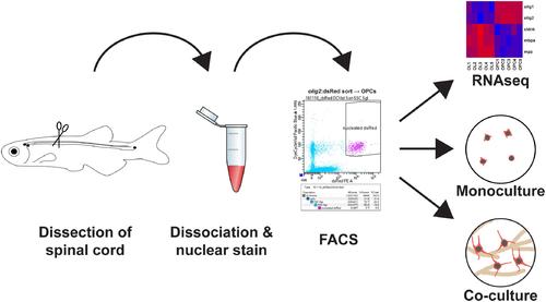
FIGURE 1. Main steps to obtain a highly pure adult zebrafish spinal oligodendrocyte progenitor cell (OPC) population: (1) Adult zebrafish spinal cord tissue is dissected. (2) The tissue is first dissociated by enzymatic digestion and then incubated with a nuclear dye. (3) The cell suspension is FACsorted based on cytoplasmic and nuclear fluorescence. (4) The purified OPCs can then be used directly for transcriptome analysis, cell culture as monoculture to study dormant OPCs or in motor neuron/OPC co-cultures to study oligodendrocyte differentiation. |
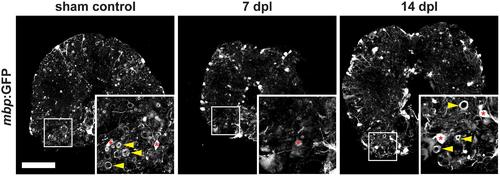
FIGURE 2. Severe demyelination and loss of oligodendrocytes (red asterisks) occur until 7 days post spinal transection lesion (dpl) in the adult zebrafish spinal cord close to the site of injury. In contrast, in sham-lesioned control spinal cords many mbp:GFP-positive myelin sheaths are found (yellow arrowheads). At 14 dpl, a partial re-establishment of the constitutive myelination pattern is observed (yellow arrowheads). 50 μm cross section; distance to lesion site ≤ 350 μm; scale bar represents 100 μm. |
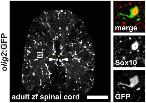
FIGURE 3. Expression of olig2:GFP in the adult zebrafish spinal cord marks OPCs in the parenchyma and a subpopulation of ventral radial glia at the central canal. OPCs are evenly distributed throughout the parenchyma in an unlesioned zebrafish spinal cord. Inset: OPCs display short arbors and co-express Sox10. 50 μm cross section; asterisk marks the central canal; scale bar represents 100 μm. |
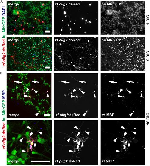
FIGURE 7. Adult zebrafish OPCs differentiate into MBP-positive mature OLs in human MN co-cultures. (A) OPCs differentiate over time in co-culture as identified by increased branching of the seeded OPCs. (B) At 16 DIC, the majority of oligodendroglial cells expresses the mature OL/myelin sheath marker protein MBP (arrowheads) while only a few cells fail to differentiate and remain MBP-negative (arrows); scale bars represent 50 μm. |
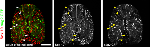
FIGURE S1 | Overlap of olig2:GFP expression and nuclear Sox10 antibody staining in the adult zebrafish spinal cord. Almost all olig2:GFP-expressing cells in the parenchyma show a nuclear signal for Sox10 (arrowheads). Also GFP-negative longitudinal structures in the parenchyma and along the midline dorsal to the central canal (asterisk) show a signal for Sox10. These structures could represent axonal processes (from sensory neurons: dorsal root and sympathetic ganglia) and meninges-like membranes. 50 μm cross section; scale bar represents 100 μm. |