- Title
-
Improving the Delivery of SOD1 Antisense Oligonucleotides to Motor Neurons Using Calcium Phosphate-Lipid Nanoparticles
- Authors
- Chen, L., Watson, C., Morsch, M., Cole, N.J., Chung, R.S., Saunders, D.N., Yerbury, J.J., Vine, K.L.
- Source
- Full text @ Front. Neurosci.
|
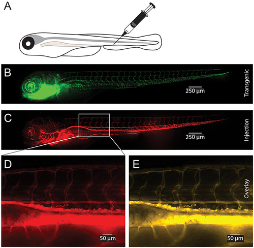
Visualization of CaP-lipid NPs after tail vein injection in transgenic zebrafish. Sterile filtered non ASO-loaded CaP-lipid NPs containing LissRdB-DSPE (stock, 1:5 or 1:10 v/v) was injected into the vein of zebrafish expressing a green fluorescent reporter in their vasculature (Tg(fli1a:EGFP)). Schematic illustration of the route of injection (A). Expression of EGFP in transgenic fish highlighting the blood vessels (B). Visualization of CaP-lipid NP in 6-day-old transgenic zebrafish 2 h after injection (C). Zoomed image of the treated zebrafish from area indicated by white box, showing distribution and accumulation of CaP-lipid NP in and around blood vessels (D). Overlay of transgenic EGFP expression and CaP-lipid NP distribution (E). EXPRESSION / LABELING:
|
|
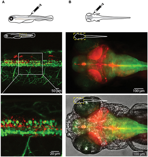
Visualization of CaP-lipid NPs and neurons in the spinal cord and brain of a 6-dpf zebrafish. Sterile filtered non ASO-loaded CaP-lipid NPs containing LissRdB-DSPE (red) were micro injected into the zebrafish spinal cord (A) and brain (B). (A) Visualization of CaP- lipid NPs containing LissRdB-DSPE (red; injections) within the spinal cord neurons (green; transgenic expression) of a 6-day-old transgenic zebrafish. Zoomed image of the treated zebrafish from area indicated by white box, showing distribution of the CaP-lipid NPs within GFP-expressing spinal cord neurons. (B) Expression of brain-injected CaP-lipid NPs (red) in a transgenic zebrafish expressing astrocyte-specific GFP [green; Tg(GFAP:GFP)] to highlight the brain-specific delivery of these particles. The bottom image overlays the bright-field channel for better visualization of the CNS. The schematic inserts in panels depict the orientation of the fish and outline the presented area. |
|
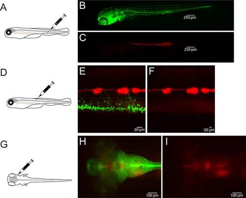
Visualization of control LissRdB injections in the zebrafish. LissRdB was injected into the vasculature (A-C), the spinal cord (D-F) and the brain (G-I) to compare distribution and brightness to CaP-lipid NPs containing LissRdB-DSPE. (A) Schematic representation of zebrafish and vein injection. (B) Transgenic zebrafish expressing EGFP (green) in the vasculature highlighting blood vessels. (C) Minimal expression of control-LissRdB throughout the vessels two hours after injection. (D) Schematic representation of zebrafish and spinal cord injection. (E) Minimal expression of control-LissRdB throughout the neurons of the spinal cord (green) two hours after injection. (F) Control-LissRdB (red) channel only. (G) Schematic representation of zebrafish and brain injection. (H) Minimal expression of control-LissRdB throughout the brain of a transgenic zebrafish (green) two hours after injection. (F) Control-LissRdB (red) channel only. EXPRESSION / LABELING:
|


