FIGURE SUMMARY
Title

Advances in the Study of Heart Development and Disease Using Zebrafish

Authors
Brown, D.R., Samsa, L.A., Qian, L., Liu, J.
Source
Full text @ J Cardiovasc Dev Dis

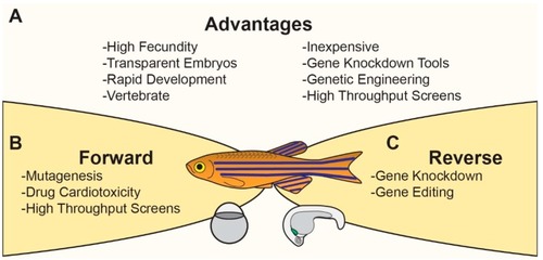
Zebrafish Model System. Schematic illustrating (A) the advantages of zebrafish as a model system, (B) forward genetic and (C) reverse genetic approaches to studying heart development and disease in zebrafish.

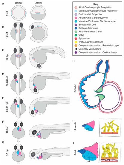
Zebrafish heart development. (AG) Lateral and dorsal views of heart development from 5 hpf embryos to 5 days post fertilization (dpf) larvae. (A) Cardiac progenitors are located at the lateral margin with the ventricular progenitors more closer to the margin than the atrial progenitors at 5 hpf; (B) Cardiac progenitors migrate bilaterally to the anterior lateral plate mesoderm by 15 hpf; (C) By 22 hpf, cardiac progenitors and developing endocardial cells have fused to form the cardiac disk which begins regular contractions between 22 and 24 hpf; (D) From 24 to 28 hpf, the disk elongates into the linear heart tube and begins leftward migration; (E) The linear heart tube continues migrating leftward and begins looping. Concurrently, from 28 to 36 hpf, second heart field cells are added to the arterial and venous poles, illustrated by shading; (F) By 48 hpf, the two chambered heart has formed; (G) The bulbous arteriosus forms at the outflow tract; (H) Cross-sectional view of the heart from 3 to 5 featuring trabeculae located primarily in the outer ventricle wall, cardiac valves, and covering of the heart by the epicardium; (I) Between larval and juvenile stages, the atrium and ventricle rotate such that the atrium is dorsal to the ventricle. The inner topology is complex and features a spongy trabecular myocardium and outer compact myocardium called the primordial layer; (J) Additional features of the adult heart are coronary arteries which feed the ventricle and expansion of the compact myocardium by addition of a cortical layer of CMs.

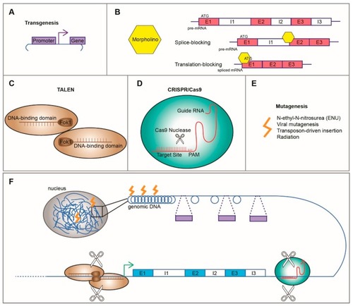
Common tools used in zebrafish include (A) transgenic insertion of foreign DNA; (B) suppression of gene expression by splice-blocking or translation-blocking morpholinos; (C) targeted genome editing by TALE-associated nucleases (TALEN); (D) targeted genome editing by CRISPR-associated Cas9 nucleases; and (E) chemical mutagenesis; (F) Schematic illustrating how these tools modify genomic DNA.

Acknowledgments
This image is the copyrighted work of the attributed author or publisher, and ZFIN has permission only to display this image to its users. Additional permissions should be obtained from the applicable author or publisher of the image. Full text @ J Cardiovasc Dev Dis