
- Title
-
Longitudinal Effects of Embryonic Exposure to Cocaine on Morphology, Cardiovascular Physiology, and Behavior in Zebrafish
- Authors
- Mersereau, E.J., Boyle, C.A., Poitra, S., Espinoza, A., Seiler, J., Longie, R., Delvo, L., Szarkowski, M., Maliske, J., Chalmers, S., Darland, D.C., Darland, T.
- Source
- Full text @ Int. J. Mol. Sci.
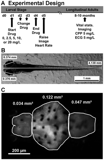
Experimental design of cocaine treatment and analysis of five-day larval zebrafish. A schematic showing the time course of embryonic drug exposure, imaging and longitudinal analysis is shown in (A); (B) shows an example of body length and eye diameter measurements made under bright field illumination; (C) shows the same fish under fluorescence illumination, focusing specifically on the brain at higher magnification. The tracing outlines the telencephalon (Tel), the diencephalon (Dien, which actually includes the optic tectum, midbrain and cerebellum), and the hindbrain (Hind, which includes the rhombencephalon), with sample area measurements given for each region. |
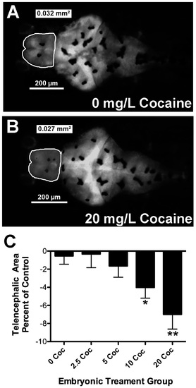
Cocaine treatment decreases telencephalic area of zebrafish larvae. There was a readily observed decrease in telencephalic size seen after treatment with the highest doses of cocaine. Panels A and B provide a comparison between untreated fish (A) and siblings treated with 20 mg/L cocaine (B); In (C), results from several experiments were combined, with brain size expressed as a percentage of untreated controls. Brain size was reduced on average 7% by treatment with 20 mg/L cocaine. Error bars signify ± SEM, * p < 0.05 relative to control and ** p < 0.01 relative to untreated control. |