- Title
-
Crypt cells are involved in kin recognition in larval zebrafish
- Authors
- Biechl, D., Tietje, K., Gerlach, G., Wullimann, M.F.
- Source
- Full text @ Sci. Rep.
|
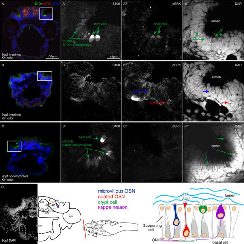
Examples of activated OSN identification and counting. All photographs shown are confocal optical sections. (A–C) Overviews of 9 dpf larval zebrafish cross-sections triple-stained for DAPI, S100 and pERK. (A'-A'''), (B'-B'''), and (C-C''') show magnified monochromatic pictures of each marker in the olfactory epithelium. Note examples of activated crypt cells in imprinted larvae tested with kin odor (A-A''') as well as some mOSNs and cOSNs (B-B'''). In non-imprinted larvae tested with kin odor, crypt cells are not activated (C-C'''). (D) shows a DAPI view of the position of the olfactory epithelium relative to eye and olfactory bulb with a corresponding explanatory drawing. Larval brain outline indicates the level of section of (D). Drawing at right bottom gives an overview on the cytoarchitectonic organization of the olfactory epithelium. Abbreviations: ac anterior commissure, CeP cerebellar plate, DT dorsal thalamus, E epiphysis, EmT eminentia thalami, H hypothalamus, Ha habenula, lG lateral glomeruli, MdG mediodorsal glomeruli, MO medulla oblongata, N region of the nucleus of the medial longitudinal fascicle, OB olfactory bulb, oc optic chiasma, ON olfactory nerve, P pallium, Po preoptic region, poc postoptic commissure, PTd dorsal part of posterior tuberculum, PTv ventral part of posterior tuberculum, S subpallium, T tegmentum, TeO tectum opticum TeVe tectal ventricle, Va valvula cerebelli, vg ventral glomeruli, VT ventral thalamus. |

