- Title
-
A K(+)-selective CNG channel orchestrates Ca2(+) signalling in zebrafish sperm
- Authors
- Fechner, S., Alvarez, L., Bönigk, W., Müller, A., Berger, T., Pascal, R., Trötschel, C., Poetsch, A., Stölting, G., Siegfried, K.R., Kremmer, E., Seifert, R., Kaupp, U.B.
- Source
- Full text @ Elife
|
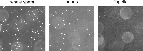
Separation of heads and flagella from whole sperm. Dark-field micrographs of whole sperm (left), purified heads (middle), and purified flagella (right). Bar represents 100 µm. |
|
Localization of the DrCNGK channel.(A) Western blot of membrane proteins (15 µg) from CHOK1 cells transfected with cDNA encoding either DrCNGK with a C-terminal HA-tag alone (lane C) or with both, a C-terminal HA-tag and an N-terminal flag-tag (N/C). Apparent molecular weight Mw is indicated on the left. (B) Characterization of anti-DrCNGK antibodies. Left: Western blot of membrane proteins (10 µg) from HEK293 cells transfected with cDNA encoding DrCNGK (Tr) and wild-type cells (wt). Right: Western blot of membrane proteins (15 µg) from zebrafish testis. (C) Western blot of membrane proteins (15 µg) from different zebrafish tissues. (D) Upper panel: Scheme of a testis cross-section. GC, germinal compartment; IC, intertubular compartment; SER, Sertoli cells; SGA, primary spermatogonia; SGB, secondary spermatogonia; SC, spermatocytes; ST, spermatids; scheme according to (Nobrega et al., 2009). Lower panel: Staining with anti-repeat1 antibody (red, left) and superposition (right) of the immunohistochemical image with a bright-field image of an in situ hybridization using an anti-DrCNGK-specific RNA probe (arrows). Bar represents 50 µm. (E) Staining of zebrafish sperm with anti-repeat1 (upper left panel) and anti-repeat3 antibody (lower left panel). Bars represent 10 µm. The respective bright-field images are shown (upper and lower right panels). (F) Western blot of equal amounts of total membrane proteins (15 µg) from purified heads and purified flagella. EXPRESSION / LABELING:
|
|
Control of loading and release of DEACM-cAMP in zebrafish sperm.(A) Dark-field micrograph (using red light) of sperm loaded with DEACM-caged cAMP (30 µM). (B) Fluorescence image after 15 s of continuous illumination with 365 nm UV light (1.75 mW power). (C) Time course of the release for the cell marked with a red circle. |
|
Sperm swimming behaviour upon Ca2+ release.(A), (B), and (C) representative swimming paths of three different DMSO loaded sperm before and after application of UV light. (D), (E), and (F) representative averaged swimming paths of three different sperm before (green) and after Ca2+ release by one (red) or two (cyan) consecutive UV flashes (black arrows). Curved blue arrows indicate the swimming direction of sperm. (G) Same swimming path shown in (F) including a temporal axis to facilitate the visualization of the changes in swimming path after consecutive flashes. Upon release (black arrows), the curvature of the swimming path progressively increases and the cell finally spins around the same position. (H) Representative flagellar shapes before (-), after Ca2+ release by one (+) or two consecutive flashes (++), and during cell spinning against the wall (bottom right). Consecutive frames every 100 ms are shown in different colours. Sequence order: red, green, blue, and yellow. (I) Mean curvature before (-) and after one (+) or two (++) UV flashes. Individual data (symbols) and mean ± sd (gray bars), number of experiments in parentheses. |


