
- Title
-
Peroxisome Proliferator-Activated Receptor γ Up-regulates Galectin-9 and Predicts Prognosis in Intestinal-Type Gastric Cancer
- Authors
- Cho, S.J., Kook, M.C., Lee, J.H., Shin, J.Y., Park, J., Bae, Y.K., Choi, I.J., Ryu, K.W., Kim, Y.W.
- Source
- Full text @ Int. J. Cancer
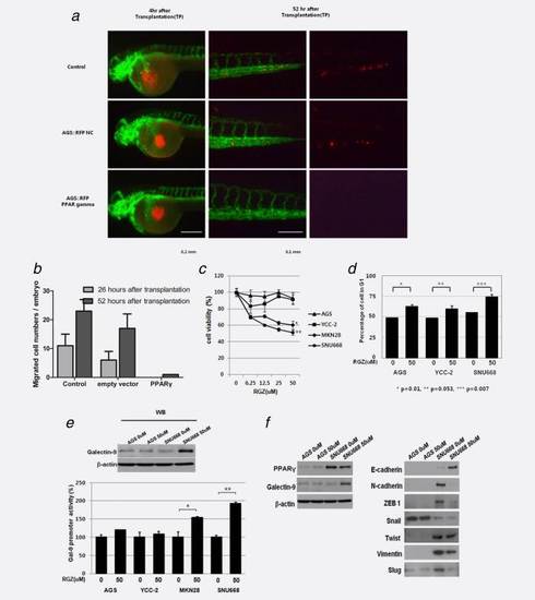
Overexpression of PPARγ blocks gastric cancer cell migration in a zebrafish xenotransplantation model, and PPARγ agonists reduce gastric cancer cell viability, induce cell cycle arrest and affect EMT. (a) At 4 hr post-transplantation (hpt), the transplanted AGS cells with red fluorescence transfected with lentivirus containing LacZ or PPARγ were located in the center of yolk sac of living transgenic zebrafish embryonic vessels are visualized with green fluorescence. 52 hpt, both control and LacZ-containing AGS cells migrated into trunk or tail region, while no PPARγ -overexpressing AGS cells had migrated to the trunk area. (b) Histogram of the number of migrated cells per embryo. (c) The effect of various concentrations of rosiglitazone on cell viability (MTT assay) of AGS, MKN28, SNU601 and NU668 cell lines. Rosiglitazone inhibited cell viability in a dose-dependent manner in MKN28 and SNU 668 cells, in which PPARγ is well expressed, but minimally affected cell viability in AGS and YCC-2 cells, in which PPARγ is poorly expressed. (d) The effect of 50 µM rosiglitazone on cell cycle of AGS, YCC-2 and SNU668 cells. *p < 0.05, **<Rosiglitazone arrested the cell cycle at G0–G1 phase in all cell lines. Cell cycle arrest in SNU 668 cells, in which PPARγ is well expressed, is more prominent than in AGS or YCC-2 cells and in which PPARγ is poorly expressed. (e) This effect occurred through galectin-9 activation; galectin-9 luciferase activity was increased after rosiglitazone treatment in MKN 28 and SNU668 cell lines (*p < 0.05 vs. control, **p < 0.01 vs. control and ***p < 0.001 vs. control). (f) Rosiglitazone (50 µM) induced the expression of galectin-9, not affecting the expression of PPARγ , increased the expression of E-cadherin and reduced the expression of N-cadherin, ZEB1, vimentin and slug in Western blotting in SNU 668 cells. |