
- Title
-
Arl13b and the non-muscle myosin heavy chain IIA are required for circular dorsal ruffle formation and cell migration
- Authors
- Casalou, C., Seixas, C., Portelinha, A., Pintado, P., Barros, M., Ramalho, J.S., Lopes, S.S., Barral, D.C.
- Source
- Full text @ J. Cell Sci.
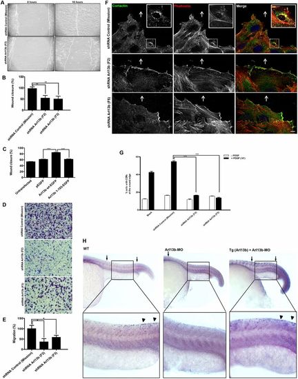
Arl13b is required for cell migration. Endogenous Arl13b was silenced by specific shRNAs (F2 and F3) in NIH-3T3 cells. (A) Control or Arl13b-silenced cells grown to confluence were scratched with a pipette tip and stimulated with PDGF to induce cell migration. Representative images taken at 0 and 16 hours post-wounding of control and Arl13b-silenced cells are shown. (B) Quantitative analysis of wound closure (%) after 16 hours was performed as described in Materials and Methods. Results are mean±s.d. of three independent experiments. (C) NIH-3T3 cells expressing Arl13b-wt–EGFP (Arl13b wt-EGFP), Arl13b-1–193–EGFP (Arl13b 1-193-EGFP) or EGFP for 24 hours were allowed to form a monolayer and then scratched. Quantitative analysis of wound closure (%) was performed 16 hours after scratching. Results are mean±s.d. of three independent experiments. (D) Arl13b-silenced or control NIH-3T3 cells were added to the upper chamber of 8-µm-pore membranes and allowed to migrate for 6 hours. Representative fields of Crystal-Violet-stained cells that migrated to the lower surface of the membranes are shown for control shRNA (Mission) and Arl13b shRNAs (F2 and F3). Scale bars: 100 µm. (E) Quantification of the transwell migration was performed as described in Materials and Methods. Results are mean±s.d. of three independent experiments. (F) Immunofluorescence analysis of CDR formation at the leading edge of migrating control and Arl13b-silenced cells. After 4 hours of incubation in the presence or absence of PDGF-BB (20 ng/ml), cells were fixed and stained with Alexa-Fluor-568-conjugated phalloidin and anti-cortactin antibody to identify CDRs. DAPI was used to stain nuclei. Arrows indicate wound direction, and insets show CDRs at higher magnification. Scale bar: 10 µm. (G) Quantification of CDRs in control (Mock and shRNA Mission) and Arl13b-silenced cells (shRNAs F2 and F5) at the edge of closing wounds. Results are mean±s.d. of three independent experiments. (H) Neural crest cell migration in Arl13b morpholino (MO)-injected zebrafish embryos. At 24 hours post fertilization, zebrafish embryos injected with Arl13b-MO and hybridized with sox10 probe, revealed that trunk neural crest cells failed to migrate (n = 17), when compared with uninjected wild-type siblings (WT, n = 18). Arrows indicate the region where neural crest cell migration occurred from anterior to posterior somites. Rescue was performed by injection of Arl13b-MO in a zebrafish transgenic line [Tg (Arl13b)], which expresses mouse Arl13b–GFP (which is not targeted by the morpholino) in addition to the endogenous zebrafish Arl13b (n = 16). Arrowheads indicate neural crest cells at the posterior somites. *P<0.05; **P<0.005 (B,E, Student's t-test); ***P<0.001 (C,G, two-way ANOVA). |