- Title
-
Wdr68 requires nuclear access for craniofacial development
- Authors
- Wang, B., Doan, D., Roman Petersen, Y., Alvarado, E., Alvarado, G., Bhandari, A., Mohanty, A., Mohanty, S., and Nissen, R.M.
- Source
- Full text @ PLoS One
|
Nuclear access is required for Wdr68 function in craniofacial development. A-H) Alcian blue stained animals. A, C, E, G) Ventral view with the Meckel′s (M), palatoquadrate (PQ) and ceratohyal (CH) labeled in red. B, D, F, H) Lateral view. A, B) Wildtype control-MO (ctrl) injected animals. C, D) Severe craniofacial phenotype of wdr68-MO injected animals co-injected with GFP mRNA (MO+GFP) that fails to rescue the craniofacial defects. E, F) Rescue of the craniofacial defects of wdr68-MO animals co-injected with GFP-Wdr68 mRNA (MO+GW). G, H) Severe craniofacial defects of wdr68-MO animals co-injected with GFPNESWdr68 mRNA (MO+GNESW) that fails to rescue the craniofacial defects. I) Graph summarizing the results of three injection trials. In aggregate, n = 50/50 (100%) for control animals, n = 47/257 (18%) for MO+GFP animals, n = 212/350 (61%) for MO+GW animals, and n = 71/385 (18%) for MO+GNESW animals. PHENOTYPE:
|
|
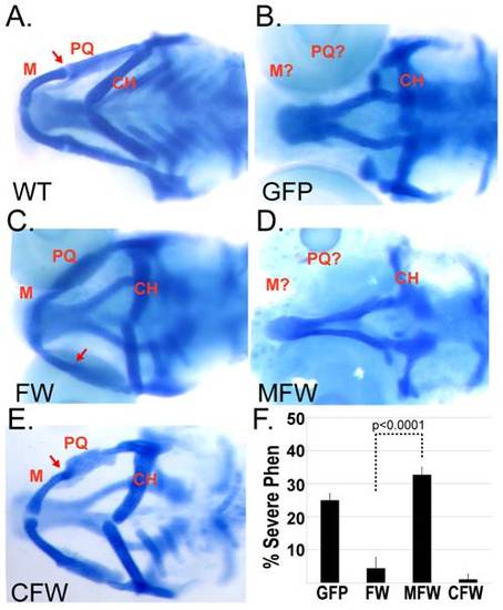
Fusion of Wdr68 to the Mad repression domain is not tolerated in vivo. A-E) Ventral views of alcian blue stained animals with the Meckel’s (M), palatoquadrate (PQ) and ceratohyal (CH) labeled in red. A) Wildtype sibling. Red arrow indicates the M-PQ jaw joint. B) Severe craniofacial defects in wdr68hi3812 animals and failure of GFP mRNA to rescue. C) Rescue of the severe craniofacial defects by injection of FlagWdr68 (FW) mRNA. Red arrow indicates the mild M-PQ joint fusion phenotype. D) Severe craniofacial defects in wdr68hi3812 animals and failure of MadFlagWdr68 (MFW) mRNA to rescue. 52/52 mutants genotyped were wdr68hi3812 homozygotes. E) Rescue of the severe craniofacial defects by injection of CebpFlagWdr68 (CFW) mRNA. Red arrow indicates the mild M-PQ joint fusion phenotype. F) Graph summarizing the results of four injection trials. In aggregate, n = 112/452 (25%) for GFP animals, n = 16/352 (4.5%) for FW animals, n = 96/296 (32%) for MFW animals, and n = 2/149 (1.3%) for CFW animals. PHENOTYPE:
|
|
The GFPNESWdr68 fusion redistributes to the cytoplasm in zebrafish. A, D) DAPI stained cell nuclei. B, E) GFP fusion proteins. C, F) Overlays of blue and green channels. A–C) Moderate nuclear enrichment of GFPWdr68 (GW) in cells of late epiboly stage zebrafish embryos. D–F) Predominant nuclear exclusion of GFPNESWdr68 (GNESW) in cells of late epiboly stage zebrafish embryos. |


