- Title
-
Sonic hedgehog is indirectly required for intraretinal axon pathfinding by regulating chemokine expression in the optic stalk
- Authors
- Hörndli C.S., and Chien, C.B.
- Source
- Full text @ Development
|
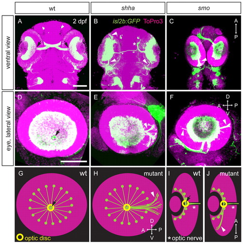
Intraretinal axon pathfinding defects in Hh pathway mutants. (A-J) Retinal projections at 2 dpf in wt, shha and smo zebrafish embryos with isl2b:GFP (green) or isl2b:tagRFP (pseudocolored green in C,F) transgene; nuclei, ToPro3 (magenta). Ventral (A-C) or lateral views (D-F) of maximum-intensity projections and schematics of wt and mutant axon projections showing lateral (G,H) and ventral views (I,J) are shown. In wt embryos (A,D,G,I), RGC axons converge at the optic disc (arrow), where they turn and pass through the optic nerve (I, asterisk). In shha (B,E,H,J) and smo (C,F,H,J) mutants, some axons fail to exit the eye, projecting posteriorly or occasionally anteriorly within the eye (arrowheads). Hh mutants also exhibit misprojections to the ipsilateral optic tectum (asterisk in B). D, dorsal; V, ventral; A, anterior; P, posterior. Scale bars: 100 μm. |
|
Hh pathway genes are expressed in the zebrafish optic stalk and RGC layer. Whole-mount in situ hybridizations (15 μm sections) for shha, ptch2 and smo mRNA. A,D,G are dorsal views; B,C,E,F,H,I are frontal views. (A) 16 hpf, shha expression in anterior midline neurectoderm (arrow). (B) 28 hpf, shha expression at the midline (arrowhead) but not optic stalk (arrow). (C) 48 hpf, shha strongly expressed at the midline (arrowhead) and in RGC layer (arrow). mRNA also detected in the optic nerve (asterisk). (D) 16 hpf, ptch2 expressed at the anterior midline (arrow). (E) 28 hpf, ptch2 strongly expressed in the optic stalk (arrow) and midline (arrowheads). (F) 48 hpf, ptch2 detected in the RGC layer (arrow) and optic nerve (asterisk). (G) 16 hpf, smo expressed throughout head region. (H) 28 hpf, smo broadly expressed, including optic stalk (arrow). (I) 48 hpf, smo localized in the RGC layer (arrow) and optic nerve (asterisk). Dashed line in A,D,G outlines the optic vesicle. A, anterior; P, posterior; D, dorsal; V, ventral; L, lateral; M, medial. Scale bar: 100 μm. |
|
Shh and Smo act non-cell-autonomously in intraretinal axon pathfinding in zebrafish. (A-G) Representative images of host eyes at 54 hpf after cell transplants at 24 hpf. Lateral views of maximum-intensity projections. Wt RGCs axons exit the eye through the optic disc (asterisk) in wt hosts (A), but often misproject (arrowheads) in shha (B) and smo (C) hosts. Shha RGC axons always exit the eye in wt hosts (D), whereas many misproject in shha hosts (E). Similar results found with smo RGCs in wt (F) or smo (G) hosts. Transplanted RGCs are isl2b:GFP (green) or isl2b:tagRFP (pseudocolored green in F,G); nuclei, ToPro3 (magenta). D, dorsal; V, ventral; A, anterior; P, posterior. Scale bar: 100 μm. (H) Percentage of embryos with misrouted donor RGC axons. Numbers of embryos shown at base of bars. *P<0.05, **P<0.01, ***P<0.001, Fisher′s exact test. |
|
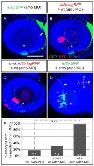
Transplants into RGC-free hosts confirm non-cell-autonomous effect of Smo in intraretinal axon pathfinding. (A-D) Maximum-intensity projections of 54 hpf isl2b:GFP or isl2b:tagRFP zebrafish embryos injected with ath5 MO at 1-cell stage (A) and transplanted with donor cells at 24 hpf (B-D). (A) No RGC differentiation in ath5 morphants; trigeminal ganglion as control for transgene expression (arrow). (B) Wt RGC axons (isl2b:tagRFP, red) in isl2b:GFP (green) ath5 morphants rarely make errors. (C) smo (isl2b:tagRFP) RGC axons in isl2b:GFP ath5 morphants are rarely misguided. (D) Wt (isl2b:GFP) RGC axons in smo ath5 morphants make errors (arrowhead). Asterisk indicates optic disc. Nuclei, ToPro3 (blue). D, dorsal; V, ventral; A, anterior; P, posterior. Scale bar: 100 μm. (E) Percentage of embryos with misrouted axons. Numbers of embryos shown at base of bars. ***P<0.005, Fisher′s exact test. |
|
Shh is required during eye patterning for intraretinal axon pathfinding in zebrafish. SANT75 (40 μM) was bath applied to inhibit Hh signaling for specific stages of embryonic development. (A,B) Whole-mount in situ hybridizations (15 μm sections), dorsal views. ptch2 mRNA is expressed at the midline (arrow) in DMSO-treated embryos (24 hpf) (B), whereas expression is lost (arrow) after SANT75 treatment (1-24 hpf) (A). (C,D) Maximum-intensity projections of lateral views (54 hpf). DMSO (1%) (D) does not affect RGC axon projections in isl2b:GFP embryos (green), whereas 40 μM SANT75 (1-54 hpf) (C) yields misguided RGC axons (arrowheads). Nuclei, ToPro3 (magenta); optic disc indicated by asterisk. (E,F) Different SANT75 application time points (E) and percentage of embryos with resulting intraretinal guidance errors (F). Number of embryos shown at base of bars. Error bars represent s.d. ne3 experiments, **P<0.01, ***P<0.001, Student′s t-test. Line drawings (E) adapted with permission (Kimmel et al., 1995). D, dorsal; V, ventral; A, anterior; P, posterior. Scale bars: 100 μm. |
|
Expression of optic stalk markers is decreased in Hh mutants. (A-C2) Maximum-intensity projections of zebrafish embryos stained for Pax2a (green) by immunohistochemistry (28 hpf); nuclei, ToPro3 (blue). Ventral views. Pax2a reduced in shha (B) and absent in smo (C) in optic stalk (arrow) compared with wt (A). Insets (A2-C2) show magnified optic stalk region, Pax2a staining only. (D-F) Whole-mount in situ hybridizations (15 μm sagittal sections; 28 hpf). netrin1a at the optic fissure (arrowheads) is decreased in shha (E) and lost in smo (F) compared with wt (D). (G-I) Coronal sections of whole mount in situ hybridizations (28 hpf). cxcl12a expression in optic stalk (arrowheads) is reduced in shha (H) and lost in smo (I) compared with wt (G). D, dorsal; V, ventral; A, anterior; P, posterior. Illustration below shows plane of views for panels above. Adapted with permission (Kimmel et al., 1995). Scale bars: 100 μm. |
|
Cxcl12a acts as an RGC axonal attractant in wt and shha and interacts genetically with the Hh pathway in zebrafish. (A-H) Maximum-intensity projections of ventral (A,C,D) and lateral (B,E-H) views at 2 dpf. (A,B) Cxcl12a mutants exhibit intraretinal axon guidance errors (arrowheads). Isl2b:GFP (green); nuclei, ToPro3 (magenta). (C) Normal axonal projections in hsp70l:EGFP embryos after heatshock. (D) Global Cxcl12a-2A-EGFP overexpression induces intraretinal axon guidance errors (arrowheads). α-tubulin (pseudocolored green); nuclei, ToPro3 (magenta), EGFP not shown. (E-H) Cxcl12a-expressing cells attract RGC axons in wt and shha embryos. Transplanted EGFP- or cxcl12a-2A-EGFP-expressing cells (green), α-tubulin (red); nuclei, ToPro3 (blue). (E) Anterior EGFP-expressing cells in wt embryos do not affect RGC outgrowth. (F) Anterior projections in wt embryos with anterior Cxcl12a-expressing cells. (F′) Substack of boxed region in F with misguided axons (red arrowhead). (G) Rare anterior projections in shha embryos with EGFP-expressing cells. (H) Shha embryos always show anterior projections with anterior Cxcl12a-expressing cells. Optic disc, asterisk. D, dorsal; V, ventral; A, anterior; P, posterior. Scale bars: 100 μm. (I) Percentage of host embryos with anterior RGC projections. Number of embryos shown at base of bars. *P<0.05, ***P<0.001, Fisher′s exact test. (J) Analysis of genetic interaction between shha and cxcl12a. Percentage of embryos per category (0-3, illustrated below graph) of RGC axon projection phenotype in shha (light gray) and shha;cxcl12a/+ (dark gray). Error bars represent s.e.m. n=3 experiments. Mann-Whitney U test, P=0.00103, of embryos ranked in four categories in shha (n=107) and shha;cxcl12a/+ (n=57) populations. |
|
Cxcr4b acts cell autonomously in RGC for correct intraretinal axon pathfinding. (A-F) Maximum-intensity projections of lateral views of 54 hpf zebrafish embryos transplanted with donor cells at 24 hpf. (A) Wt RGC axons (isl2b:GFP) in wt embryos exit the eye (asterisk). (B) Wt RGC axons (isl2b:tagRFP) often make errors in cxcr4b hosts (arrowhead). (C) cxcr4b RGC axons (isl2b:GFP) exit the eye in most cases when transplanted into wt hosts (isl2b:tagRFP). (D) Cxcr4b RGC axons (isl2b:GFP) are misguided in ath5 morphants (isl2b:tagRFP) in almost all transplants (arrowhead). (E,F) Wt RGC axons (isl2b:tagRFP) rarely make errors in ath5 morphants (isl2b:tagRFP) (E)and in cxcr4b ath5 morphants (isl2b:GFP) (F). D, dorsal; V, ventral; A, anterior; P, posterior. Scale bar: 100 μm. (G) Percentage of host embryos with misguided donor RGC axons. Number of embryos shown at base of bars. **P<0.01, ***P<0.001, Fisher′s exact test. |
|
Focal dye injections show prevalence for posterior RGC axon projections in shha mutants. Maximum-intensity confocal projections of lateral views of focal lipophilic dye injections into the retina of 2 dpf wt and shha embryos. (A) Wt eye injected with DiI anterior and DiO dorsal shows axon projections towards the optic disc (white asterisk), where the axons leave the eye through the optic nerve. (B) Diagram showing shha RGC axon projections as seen after DiI injections of 13 embryos. Axon bundles from all quadrants of the retina project towards the optic disc but fail to leave through the optic nerve and instead continue growing inside the eye. Posterior RGC projections are seen more commonly than anterior projections. (C,D) Examples of shha eyes focally injected with DiI. Misprojecting RGC axon bundles grow towards the optic disc (red asterisk) before projecting posteriorly or anteriorly (arrowheads). D, dorsal; V, ventral; A, anterior; P, posterior. |
|
Normal intraretinal axon pathfinding in netrin1a morphants. (A) Reverse-transcription PCR showing knockdown of properly spliced netrin1a mRNA in netrin1aSBMO and p53MO co-injected embryos compared with p53MO control-injected embryos. Middle band, unspliced pre-mRNA levels are increased in netrin1aSBMO-injected embryos compared with control. Bottom band, beta-actin mRNA levels serve as loading control. (B,C) Maximum-intensity confocal projections of lateral views at 2 dpf. netrin1aSBMO and p53MO co-injected embryos (B) and p53 control morphants (C) show normal intraretinal axon pathfinding, whereas eye size is decreased in netrin1a morphants compared with control embryos. Photoreceptor differentiation is visible in both netrin1a morphants and controls (arrows). isl2b:GFP (green); nuclei, ToPro3 (magenta). Scale bar: 100 μm. |


