
- Title
-
Close association of olfactory placode precursors and cranial neural crest cells does not predestine cell mixing
- Authors
- Harden, M.V., Pereiro, L., Ramialison, M., Wittbrodt, J., Prasad, M.K., McCallion, A.S., and Whitlock, K.E.
- Source
- Full text @ Dev. Dyn.

Neural crest (sox10) and olfactory placode (six4b, dlx3b) markers have distinct but partially overlapping expression patterns. A–D:sox10 in situ hybridization (A,C) and anti-green fluorescent protein (GFP) immunocytochemistry (B,D) in sox10:EGFP embryos. At 5s (somite stage; A,B), sox10 mRNA expression (A) is similar to GFP expression (B), but at 14s–15s (C,D), GFP (D) is expressed in more cells than is sox10 mRNA (C). E,F: Double labeling of the olfactory placode (OP) field and cranial neural crest cells (CNCCs) in sox10:EGFP embryos at 2s. six4b (E, purple) and dlx3b (F, blue) are expressed in the OP field at 2s. Both genes are expressed in a horseshoe shape around rostral neural plate. six4b is more restricted than dlx3b. At this stage, the premigratory CNCCs (E,F, brown) flank the posterior domains of the six4b (E, purple) and dlx3b (F, blue) fields. A–D: Lateral view, rostral to the right, dorsal to the top of the page. E,F: Dorsal views, rostral to the top of the page. hpf, hour postfertilization. Scale bars = 100 μm in A–D, 100 μm in E,F. |

dlx3b-expressing olfactory placode (OP) precursors share a common border with cranial neural crest cells (CNCCs). Visualization of OP convergence (using dlx3b in situ hybridization, in blue) and the CNC field (using anti-green fluorescent protein [GFP] immunocytochemistry, in brown), in sox10:EGFP embryos. A–I: Dorsal views, rostral to the top of the page, of fixed-staged, double-labeled embryos. Embryos were examined every 2 somites. E,E′: Two different focal planes of the same embryo at 12s (somite stage): E, dorsal olfactory placode; E′, ventral edge of the olfactory placode. F,F′: Two different focal planes at 14s: F, dorsal olfactory placode; F′, ventral edge of the olfactory placode. The CNCCs are both dorsal and ventral to the olfactory fields at these stages. J,K: Ventral views (rostral to the top) of the formed olfactory placode surrounded by CNCCs at 18s (J) and 20s (K). Scale bars = 30 μm in A (applies in A–K). Twenty embryos were examined per time point. |
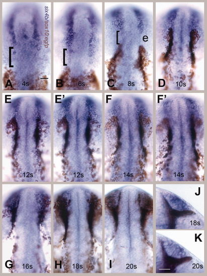
six4b-expressing olfactory placode (OP) precursors do not share a common border with cranial neural crest cells (CNCCs). Visualization of OP field using six4b (in blue) and CNCCs using anti-green fluorescent protein (GFP) immunocytochemistry (in brown) in sox10:EGFP embryos. A–I: Dorsal views, rostral to the top of the page. E,E′: Two different focal planes of same embryo at 12 somites: E, dorsal OP; E′, ventral edge of the OP. F,F′: Two different focal planes at 14s (somite stage): F dorsal OP, F′ ventral edge of the OP. The neural crest cells are both dorsal and ventral to the olfactory fields at these stages. G–I: CNCCs move dorsally over the forming OP. J,K: Ventral views of the formed OP surrounded by CNCCS at 18s (J) and 20s (K), rostral to top. Scale bars = 30 μm in A (applies in A–K). Twenty embryos were examined per time point. |
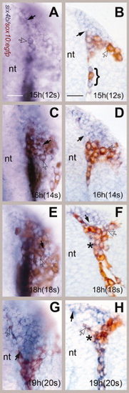
Cranial neural crest cells (CNCCs) move rostrally surrounding the forming olfactory placode (OP). A–H: Whole-mount preparations (A,C,E,G) and cryostat sections (B,D,F,H) of six4b (in situ, blue)/ anti- green fluorescent protein (GFP; immunocytochemistry, brown) double-labeled embryos. All images are dorsal views, rostral to top of the page. A,B: At 12s (somite stage), the neural crest cells were first seen meeting the posterior edge of the OP with some mixing of CNCCs and OP cells (B, bracket). C,D: At 14s, the CNCCs cells began to aggregate at the posterior border of the OP. E,F: At 18s, the neural crest cells surrounded the OP. G,H: The border of the OP was refined at 20s. six4b-expressing cells (black arrows) were observed at the edge of the forming OP. E,F: Some cells (asterisks) appeared to be double labeled. nt, neural tube. Scale bars A (for A–G) and B (for B–H) = 30 μm. Five whole-mount and sectioned embryos were examined at high magnification per time point. |
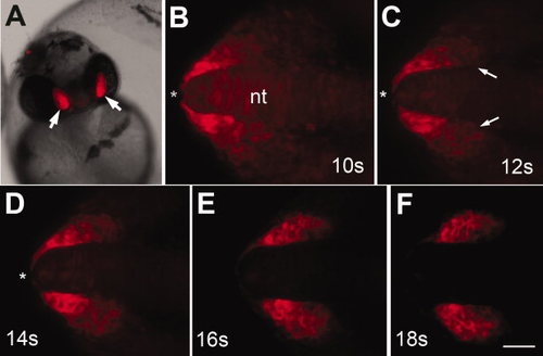
Six4b:mCherry expression starts during somitogenesis. A: Expression in olfactory placodes (OPs) in founder at 48 hours postfertilization (hpf). B–F: Stills from 6-hr time lapse generated starting at 10s (see Supp. Movie S1). B–F: The OP precursors are concentrated in the most rostral region of the head (asterisk). C–F: The mCherry-expressing cells located at the posterior edge of the OP field (C, arrows) coalesce, forming the posterior border (D–F). B–D: During this time, the anterior mCherry-expressing cells move caudally away from the tip of the neural tube (asterisks) aggregating to form the olfactory placodes (E,F). All images are dorsal views with rostral to the left. nt, neural tube. Scale bar = 30. (See Supp. Movie S2, S3.) |
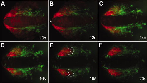
Neural crest cells migrate with olfactory placode precursors. Images from time lapse movie showing migration of cranial neural crest cells (CNCCs; green) and olfactory placode (OP) precursors (red). A: At 10s (somite stage), OP precursors (red) are wrapped around the rostral end of the neural tube (asterisk). B: At 12s, the OP precursors move away from the rostral midline (asterisk) as CNCCs (green) advance rostrally. C: By 14s, the CNCCs start to surround the posterior edge of the OP precursors. D: The OP precursors (red) are no longer evident at the tip of the neural tube at 16s. CNCCs (green) arrive at the anterior limit of the OPs. E: At 18s, the CNCCs (green) have migrated to the rostral limit of the olfactory placodes. The posterior region of the OP contains neither green nor red cells (white outline), which is the presumed region of dlx3b expression. F: At the end of the movie (20s), the olfactory placodes are fully formed. All images are dorsal views with rostral to the left. Scale bar = 30 μm (see S3, S4 for movies). |

Six4b:mCherry is not expressed in differentiated sensory neuron subtypes in the olfactory epithelia. A–C: TRPC2:Venus-expressing microvillar sensory neurons in the olfactory organ at 5 days postfertilization (dpf). A,C: The TRPC2:Venus and six4b:mCherry signals do not colocalize (arrow); the TRPC2:Venus-expressing neurons lie apical to the six4b:mCherry-expressing cells (A,C, red). D–F: OMP:YFP-expressing ciliated sensory neurons (green) of the olfactory organ are widely distributed throughout the olfactory epithelia (D) but do not express six4b:mCherry (bracket). E,F: six4b:mCherry-expressing cells appear to surround the sensory neurons with the signal of the sensory neurons (E, bracket) localized to the nonexpressing region of the mCherry (F, bracket). All pictures are anterodorsal views. Scale bar = 25 μm. |