- Title
-
Conserved Function of lincRNAs in Vertebrate Embryonic Development despite Rapid Sequence Evolution
- Authors
- Ulitsky, I., Shkumatava, A., Jan, C.H., Sive, H., and Bartel, D.P.
- Source
- Full text @ Cell
|
Expression of Zebrafish lincRNAs (A) Whole-mount in situ hybridizations of selected lincRNAs. Control experiments using sense probes for selected lincRNAs were also performed (Figure S2). (B) Expression levels of lincRNA and protein-coding genes evaluated using RNA-Seq results from ten stages/tissues. Plots indicate the median, quartiles, and 10th and 90th percentiles. RPKM is reads per kilobase per million reads. (C) Correlations between levels of neighboring transcripts. For each gene, the Spearman correlation between its expression profile (across ten stages/tissues) and that of the closest protein-coding gene was determined, and the average is plotted for the lincRNA and coding genes. Error bars are 95% confidence intervals based on 1,000 random shuffles of lincRNA positions. See also Figure S2 and Table S4. EXPRESSION / LABELING:
|
|
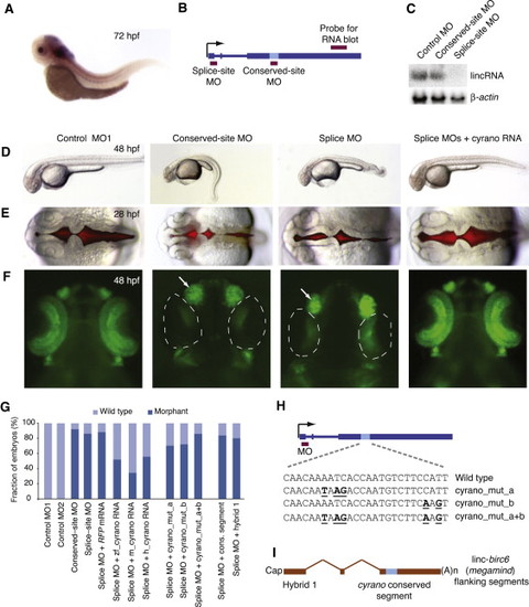
The Importance of linc-oip5 for Proper Embryonic Development (A) In situ hybridization showing cyrano expression in the CNS and notochord of zebrafish embryos at 72 hpf. (B) Gene architecture of cyrano, showing the hybridization sites of the RNA-blot probe and MOs (red boxes). (C) RNA blot monitoring cyrano accumulation in wild-type embryos (48 hpf) that had been injected with the indicated MOs. To control for loading, the blot was reprobed for β-actin mRNA. (D) Embryos at 48 hpf that had been either injected with the indicated MO or coinjected with the splice-site MO and mature mouse cyrano RNA. (E) Brain ventricles after injection with the indicated reagents, visualized using a red fluorescent dye injected into the ventricle space at 28 hpf. (F) Embryos at 48 hpf that had been injected with the indicated reagents. NeuroD-positive neurons in the retina and nasal placode were marked with GFP expressed from the neurod promoter (Obholzer et al., 2008). Near absence of NeuroD-positive neurons in the retina (dotted line) and enlargement of the nasal placode (arrow) are indicated.n (G) Frequency of morphant phenotypes in injected embryos (Table S5). (H) Schematic of DNA point substitutions in the cyrano-conserved site. (I) Architecture of a hybrid transcript containing the cyrano-conserved segment in the context of linc-birc6 (megamind)-flanking sequences. See also Figure S4 and Table S5. EXPRESSION / LABELING:
PHENOTYPE:
|
|
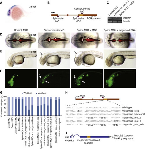
The Importance of linc-birc6 for Proper Brain Development (A) In situ hybridization showing megamind expression in the brain and eyes of zebrafish embryos at 28 hpf. (B) Gene architecture of megamind, showing the hybridization sites of the MOs (red boxes) and RT-PCR primers (arrows). (C) Semiquantitative RT-PCR of mature megamind in embryos at 72 hpf that had been injected with the indicated MOs. β-actin mRNA was used as a control. (D) Brain ventricles after injection with either the indicated MOs or coinjected with the splice-site MO and mature mouse megamind RNA, visualized using a red fluorescent dye injected into the ventricle space at 28 hpf. An expanded midbrain ventricle (arrow) and abnormal hindbrain hinge point (asterisk) are indicated. (E) Embryos at 48 hpf that had been injected with the indicated reagents. Abnormal head shape and enlarged brain ventricles are indicated (arrow). (F) Embryos at 48 hpf that had been injected with the indicated reagents. NeuroD-positive neurons in the retina and nasal placode were marked with GFP expressed from the neurod promoter (Obholzer et al., 2008). Near absence of NeuroD-positive neurons in the retina and tectum (arrows) is indicated. (G) Frequency of morphant phenotypes in injected embryos (Table S5). (H) Schematic of DNA point substitutions in the megamind-conserved segment. (I) Architecture of a hybrid transcript containing the megamind-conserved segment in the context of cyrano-flanking sequences. See also Figure S5 and Table S5. |
|
Whole-Mount In Situ Hybridizations Using Sense Probes for Selected lincRNAs, Related to Figure 2. |
|
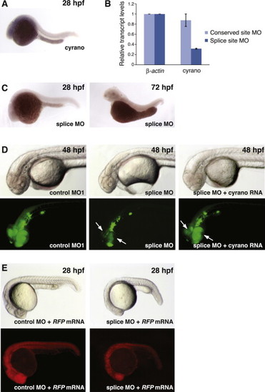
Characterization of linc-oip5 (cyrano) and NeuroD Expression in Zebrafish Embryos, Related to Figure 5 (A) In situ hybridization showing cyrano expression in the brain and notochord of zebrafish embryos at 28 hpf. (B) Reduced cyrano levels in embryos injected with the cyrano splice-site MO measured by qRT-PCR. (C) In situ hybridization of cyrano in embryos injected with the cyrano splice-site MO. (D) Embryos at 48 hpf that had been injected with the indicated reagents. Bottom panel shows subsets of neurons expressing GFP driven by the neurod promoter. Arrows point at NeuroD-positive neurons in the retina and tectum at 48 hpf. (E) Embryo coinjected with mock RNA (RFP) and the indicated MOs. Bottom panel shows RFP expression. |
|
Characterization of linc-birc6 Expression in Zebrafish Embryos, Related to Figure 6 (A) In situ hybridization showing megamind expression in the brain of zebrafish embryos at 72 hpf. (B) Reduced megamind levels in embryos injected with the megamind splice-site MOs measured by qRT-PCR. (C) In situ hybridization of megamind expression in zebrafish embryos injected with the splice site MOs. (D) Embryo at 28 hpf that had been injected with the indicated reagents. Bottom panel shows RFP expression. Arrow points at defects in brain development. EXPRESSION / LABELING:
|

ZFIN is incorporating published figure images and captions as part of an ongoing project. Figures from some publications have not yet been curated, or are not available for display because of copyright restrictions. EXPRESSION / LABELING:
|
Reprinted from Cell, 147(7), Ulitsky, I., Shkumatava, A., Jan, C.H., Sive, H., and Bartel, D.P., Conserved Function of lincRNAs in Vertebrate Embryonic Development despite Rapid Sequence Evolution, 1537-1550, Copyright (2011) with permission from Elsevier. Full text @ Cell


