- Title
-
A zebrafish SKIV2L2-enhancer trap line provides a useful tool for the study of peripheral sensory circuit development
- Authors
- Cox, J.A., McAdow, A.R., Dinitz, A.E., McCallion, A.S., Johnson, S.L., and Voigt, M.M.
- Source
- Full text @ Gene Expr. Patterns
|
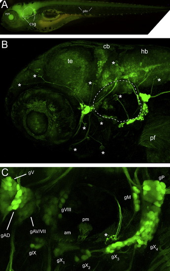
GFP expression of SKIV2L2 enhancer trap line. (A) Whole mount epifluorescence image of Tg(SKIV2L2:gfp) at 4 days- note clusters of GFP+ cells surrounding otic vesicle (dotted line). (B) Confocal microscopy of GFP expression in the peripheral ganglia in the head at 4 days. Asterisks represent neuromasts. Te, telencephalon, cb, cerebellum, hb, hindbrain. (C) A detailed view of the sensory ganglia labeled by the SKIV2L2 enhancer. Am, anterior macula; gAD, dorsal anterior lateral line ganglia; gAV, ventral anterior lateral line ganglia; gV, trigeminal ganglia; gVII, facial ganglia; gVIII, statoacoustic ganglia; gIX, glossopharyngeal ganglia, gX1-4, vagal ganglia, gM, medial lateral line ganglia, gP, posterior lateral lined ganglia; plln, posterior lateral line; pm, posterior macula; *neuromast. EXPRESSION / LABELING:
|
|
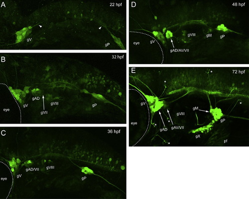
Developmental expression of Tg(SKIV2L2:gfp) in the head. (A) 22 hpf. Trigeminal and posterior lateral line ganglia are labeled. (B) 32 hpf. The dorsal anterior lateral line ganglia and the statoacoustic ganglia have appeared. (C) 36 hpf. (D) 48 hpf. The ventral anterior lateral line ganglia and the medial lateral line ganglia have developed. (E) 60 hpf. All the ganglia that are labeled at 4 days are present and will coalesce and send out more processes over the 30 h to form the pattern seen in Fig 1. gAD, dorsal anterior lateral line ganglia; gAV, ventral anterior lateral line ganglia; gV, trigeminal ganglia; gVII, facial ganglia; gVIII, statoacoustic ganglia; gIX, glossopharyngeal ganglia, gX1-4, vagal ganglia, gM, medial lateral line ganglia, gP, posterior lateral lined ganglia; *neuromast Views are lateral, anterior to the left. EXPRESSION / LABELING:
|
|
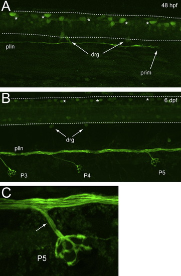
Developmental expression of Tg(SKIV2L2:gfp) in the trunk. (A) 48 hpf. GFP-labeled Rohon-Beard neurons (asterisks) can be seen in the spinal cord. Some dorsal root ganglia neurons (drg) are labeled at the dorsal side of the spinal cord. The posterior lateral line (plln) can be seen growing caudally (prim, lateral line primordium). (B) 6 days. Rohon-Beard neurons are present (asterisks) and the plln has separated into distinct fascicles and can be seen to send off axonal branches to the neuromasts (P3, P4, P5). The drg neurons are more distinct. (C) A higher power image of neuromast P5 showing multiple axon fascicles (arrow) leaving the plln to innervate the neuromast. |
|
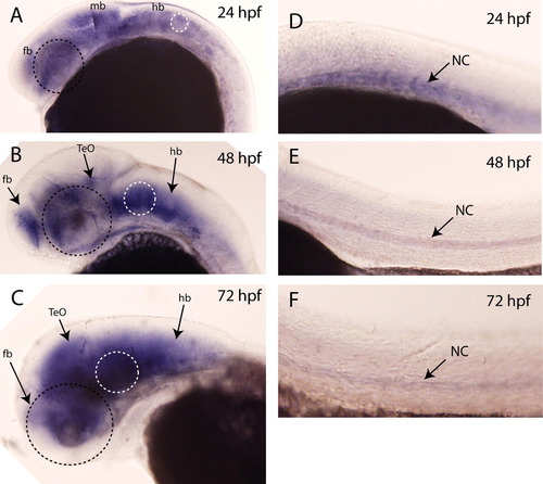
Expression pattern of lhfpl4 by whole mount in situ hybridization. (A) At 24 hpf, lhfpl4 is expressed throughout the brain (fb, forebrain, mb, midbrain, hb, hindbrain). At 48 hpf (B) and 72 hpf, (C) its expression is still throughout the brain, especially in the forebrain, the optic tectum (TeO) and in the hindbrain. Expression in the trunk (D–F) is restricted to the notochord (nc). There is no staining evident in any peripheral ganglia, in the spinal cord or in the lateral line. Black dotted circle indicates position of the eye; white dotted circle indicates position of the ear. EXPRESSION / LABELING:
|
Reprinted from Gene expression patterns : GEP, 11(7), Cox, J.A., McAdow, A.R., Dinitz, A.E., McCallion, A.S., Johnson, S.L., and Voigt, M.M., A zebrafish SKIV2L2-enhancer trap line provides a useful tool for the study of peripheral sensory circuit development, 409-14, Copyright (2011) with permission from Elsevier. Full text @ Gene Expr. Patterns


