
- Title
-
A zebrafish melanophore model of amyloid beta toxicity
- Authors
- Newman, M., Wilson, L., Camp, E., Verdile, G., Martins, R., and Lardelli, M.
- Source
- Full text @ Zebrafish
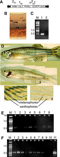
(A) The pT2-mitfa-Aβ-EGFP transgene. The transposon construct in the Sleeping Beauty vector. The binding positions of the primer sets used for polymerase chain reaction (PCR) tests are indicated above the transgene. Arrows at the end of the transgene are the inserted repeats recognized by the transposase mRNA. (B) Whole-mount in situ transcript hybridization for transcripts containing EGFP coding sequences in a 24 hpf embryo. A lateral view (slightly oblique, sagittal optical section) at the midregion of the yolk extension (ye). Dorsal is to the top. Arrowheads indicate putative immature melanophores lacking pigmentation. The upper indicated cell was observed to lie near the level of the horizontal myoseptum in the region between the notochord (nc) and the myotome (my), consistent with the medial pathway of melanocyte migration in zebrafish ( Jesuthasan, 1996). The indicated cells appeared somewhat dendritic. (C) Nested reverse transcription (RT)-PCR to detect mitfa transcription in the skin of wild-type adult zebrafish. Lane 1 is the PCR product from wild-type skin; lane 2 is the negative (no RNA/cDNA) control. M indicates the DNA marker (‘‘2-Log DNA Ladder’’). (D) Adult zebrafish at 16 months. Upper panel: a representative normal adult zebrafish. Lower panel: a transgenic fish that had specific loss of melanophores. (1) and (2) are enlarged images of corresponding areas of the caudal fin (see boxes). (E, F) PCR on genomic DNA from zebrafish tail clips to detect the presence of the transgene. Eight striped fish (E, lanes 1–8) and eight stripeless fish (F, lanes 1–8) from an outcross of line 1 were tail clipped, and genomic DNA was tested for the presence of the transgene. ‘+’ indicates detection of transgene and ‘-’ indicates no detection. (F) Lane 9 is blank, lane 10 is a negative control (no DNA) reaction, and lane 11 is from a reaction containing pT2-mitfa-Aβ-EGFP plasmid DNA as a template as a positive control. M indicates the DNA marker, which is ‘‘2-Log DNA Ladder’’ supplied by New England BioLabs (Ipswich, MA). Color images available online at www.liebertonline.com=zeb. |

Unillustrated author statements PHENOTYPE:
|