
- Title
-
Let It Flow: Morpholino Knockdown in Zebrafish Embryos Reveals a Pro-Angiogenic Effect of the Metalloprotease Meprin alpha(2)
- Authors
- Schütte, A., Hedrich, J., Stöcker, W., and Becker-Pauly, C.
- Source
- Full text @ PLoS One
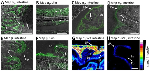
Distribution of meprin α1, α2 and β in zebrafish tissues. Immunofluorescence microscopy of cryosections from whole mount zebrafish, using specific peptide antibodies, revealed meprin α1 in the brush border cells of intestinal epithelia (Ep) (A) and epidermis (B), whereas meprin α2 was observed in the lamina propria mucosae (LP) only (C, D). Additionally, meprin β signals could be detected in the zebrafish intestine (Ep) and epidermis (Ed) (E, F respectively). To verify the efficiency of meprin knockdowns due to morpholino injection, we compared the fluorescence signal intensity in cryosections of wild type (G) and meprin α2 deficient embryos (H). Evidently, the expression of meprin α2 in the lamina propria (LP) of the intestine is largely decreased (G, H). Similar analyses of the meprin β morphant were not possible, due to the lethality within 24 hpf. (Ep: epithelium; Lu: lumen; LP: lamina propria; Ed: epidermis; De: dermis; scale bars: 25 μm; signal intensity was calculated with ImageJ V.1.41o). EXPRESSION / LABELING:
|
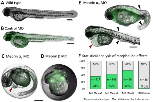
Morpholino knockdown in zebrafish embryos exhibit severe phenotypes of meprin α1, α2 and β (C–E). Wild type (A, 60 hpf) and control fish (B, 42 hpf) did not show any defects in embryonic morphology and development. In meprin α1 morphants (C, 32 hpf), only slight defects like the dilation of pericard (red arrow) were visible, whereas meprin β morphants (D, 22 hpf) showed striking tissue disorganization in trunk and tail, leading to early death within 24 hpf. Embryos injected with morpholinos against meprin α2 (E, 42 hpf), likewise exhibited dilated pericards (red arrow), but also showed severe epidermal abnormalities in trunk and tail (black arrows). Statistical analyses visualize the frequency of morphant phenotypes (F). ‘n’ describes the number of injected embryos. Morpholinos were tagged with carboxyfluorescein (green fluorescence). (Scale bars: 250 μm). PHENOTYPE:
|
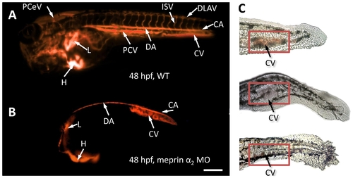
The vascular system of meprin α2 knockdown embryos exhibits dramatic defects (B, C). Microangiography (with TRITC-Dextran) revealed a drastically reduced vascular system without any intersegmental blood vessels (B) in meprin α2 morphants, compared to the non-injected wild type zebrafish (A). Additionally, erythrocytes accumulated in the ventral caudal tail region (C), possibly as a consequence of this sprouting failure. (PCeV: Posterior cerebral vein; ISV: intersegmental vessels; DLAV: dorsal longitudinal anastomotic vessel; CA: caudal artery; DA: dorsal aorta CV: caudal vein; PCV: posterior cardinal vein; L: liver; H: heart) (Scale bar: 250 μm). PHENOTYPE:
|
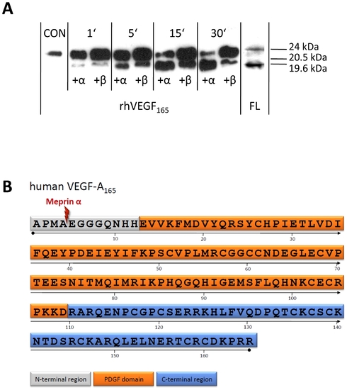
Human meprin α and β are capable of processing VEGF-A specifically. (A) The cleavage of recombinant human VEGF-A165 (CON, untreated, 24 kDa) by recombinant meprin α and β (each 85 nM) for 1 to 30 min resulted in two fragments of different molecular weight (19.6 kDa in case of meprin α, 20.5 kDa by meprin β), visualized by western blot analysis. In wild type zebrafish whole lysates (FL), VEGF-A could be detected using specific antibodies indicating a fragment similar to that produced by meprin processing. (B) Domain structure of human VEGF-A165 (P15692-4). The lightning indicates the cleavage site between Ala4 and Glu5, identified after incubation with recombinant human meprin α. PDGF (platelet-derived growth factor). In addition, the N-terminal cleavage site in human VEGF-A165 between Ala30 and Glu31 due to recombinant human meprin α activity could be identified by N-terminal sequencing. EXPRESSION / LABELING:
|

Unillustrated author statements PHENOTYPE:
|