
- Title
-
Introduction of a Foreign Gene into Zebrafish and Medaka Cells Using Adenoviral Vectors
- Authors
- Kawasaki, T., Saito, K., Mitsui, K., Ikawa, M., Yamashita, M., Taniguchi, Y., Takeda, S., Mitani, K., and Sakai, N.
- Source
- Full text @ Zebrafish
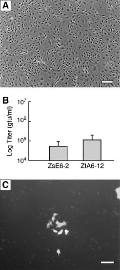
Infection efficiency of the lentiviral vector in zebrafish cells. (A) Morphology of ZsE6-2 cells. Scale bar, 200 μm. (B) Infection efficiency of lentiviruses in ZsE6-2 and ZtA6-12 cells. The vertical bar indicates the SD of duplicate independent experiments in which treatments were performed in triplicate. (C) Proliferation of green fluorescent protein–positive ZtA6-12 cells 4 days after infection. Scale bar, 100 μm. SD, standard deviation. |

Infection efficiency of the adenoviral vector in medaka Mtp2 cells. (A) Morphology of Mtp2 cells. Scale bar, 200 μm. (B) Infection efficiency of Ads in Mtp2 cells. (C) Fold increases in the infection efficiency of Ads via the use of magnetic particles. The vertical bar indicates the SD of duplicate independent experiments in which treatments were performed in triplicate. |

Infection of zebrafish male germ cells with the adenovirus vector. (A–C) Infected spermatogonia (arrow head). (D–F) An infected primary spermatocyte (or spermatogonium) in the clump (arrow head). (G–I) Infected secondary spermatocytes (arrow head). (A, D, G) Bright field, (B, E, H) dark field, and (C, F, I) merged fields. Scale bar, 100 μm. |