
- Title
-
Bone growth in zebrafish fins occurs via multiple pulses of cell proliferation
- Authors
- Jain, I., Stroka, C., Yan, J., Huang, W.M., and Iovine, M.K.
- Source
- Full text @ Dev. Dyn.
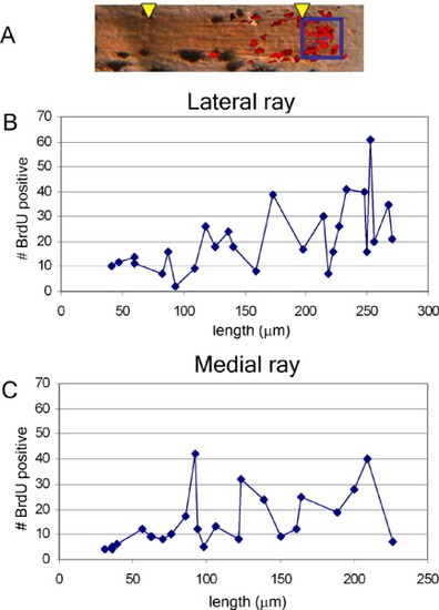
Cell proliferation during segment growth appears pulsatile. A: Overlayed images of ZNS5+ cells (osteoblasts) in brown and bromodeoxyuridine-positive (BrdU+) cells in red. Arrowheads indicate joints. BrdU+ cells distal to the most distal joint were counted (boxed region) from individual fin rays and plotted against the length of the ultimate segment from the same fin. B: Number of BrdU+ cells in the longest fin ray in the ventral lobe. C: Number of BrdU+ cells in the shortest fin ray in the fin. Each data point in B and C represents a single fin ray. N = 28 rays. |
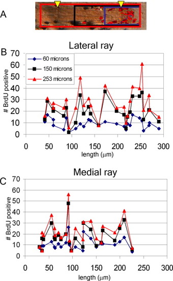
Cells proximal to the most distal joint appear to contribute to segment growth. A: Overlayed images of ZNS5+ cells (osteoblasts) in brown and bromodeoxyuridine-positive (BrdU+) cells in red. Arrowheads indicate joints. BrdU+ cells were counted from three areas: 60 μm from the end of the fin (blue box), 150 μm from the end of the fin (black box), 250 μm from the end of the fin (red box); and plotted against the length of the ultimate segment in B and C. B: BrdU+ cells counted in each of the three areas, from the longest fin ray in the ventral lobe. C: BrdU+ cells counted in each of the three areas, from the shortest fin ray in the fin. Each data point in B and C represents a single fin ray. N = 28 rays. |
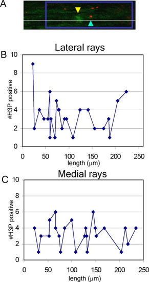
The number of mitotic cells during segment growth appears pulsatile. A: Confocal image of a single section with ZNS5+ cells in green and H3P+ cells in red. The yellow arrowhead indicates the joint; the blue arrowhead indicates a single H3P+ cell in this section (and in this fin ray). The boxed region represents an area 250 μm from the end of the fin. B: H3P + cells in the longest fin ray in the ventral lobe. C: H3P+ cells from the shortest fin ray in the fin. Each data point in B and C represents a single fin ray. N = 25 rays. |
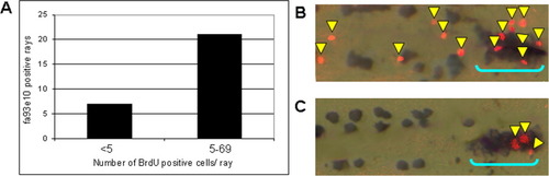
Double labeling of young fins with the growth marker fa93e10, and with bromodeoxyuridine (BrdU). A: Fins positive for fa93e10 may have a low number of BrdU-positive cells (<5; n = 7) or a high number of BrdU-positive cells (≥ 5; n = 21). B: An example of a fa93e10-positive fin (bracket) with a high number of BrdU-positive cells (arrows, n = 13). C: An example of a fa93e10-positive fin (bracket) with a low number of BrdU-positive cells (arrows, n=3). Brown cells are melanocytes. N = 28 fins. EXPRESSION / LABELING:
|