
- Title
-
Ubc9 interacts with a nuclear localization signal and mediates nuclear localization of the paired-like homeobox protein Vsx-1 independent of SUMO-1 modification
- Authors
- Kurtzman, A.L. and Schechter, N.
- Source
- Full text @ Proc. Natl. Acad. Sci. USA
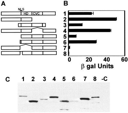
zUbc9 interacts with a NLS at the NH2 terminus of the homeodomain. (A) Vsx-1 deletion mutants: 1, zVsx-1(ORF); 2, zVsx-1ΔC-term; 3, goldfish Vsx-1 target; 4, zVsx-1ΔCVC; 5, zVsx-1ΔN-term1; 6, zVsx-1ΔN-term2; 7, zVsx-1ΔNLS/HD; and 8, zVsx-1ΔNLS. (B) Liquid culture assay using CPRG as substrate (CLONTECH) was used to calculate β-galactosidase activity. One unit of β-galactosidase equals the amount that hydrolyzes 1 μmol of CPRG to chlorophenol red and D-galactose per minute per cell. In the graph, the means (±SEM) are the result of samples processed in triplicate. (C) Anti-LexA antibody Western blot of yeast lysates shows expression of the LexA- binding domain fusion proteins noted in A. |

Zebrafish Vsx-1 is not a substrate for SUMO-1 conjugation in vitro or in COS-7 cells. Open and closed arrows indicate unconjugated MT-zVsx-1 and RanGAP1, respectively. Asterisk shows RanGAP1 conjugated to endogenous SUMO-1. (A) Xenopus RanGAP1 (lanes 1-4) or MT-zVsx-1 (lanes 6-9) were in vitro translated and labeled with [35S]Met/[35S]Cys. Recombinant GST-SUMO-1 and Ubc9 were added as indicated. Ubc9 (3x) was added to lanes 4, 5, 9, and 10. Western blots using anti-SUMO-1 or anti-hsUbc9 antibodies show relative levels of GST-SUMO-1 and Ubc9. (B) COS-7 cells were transfected with expression plasmids encoding RanGAP1, MT-zVsx-1, and GFP-SUMO-1. Cell lysates were immunoblotted with anti-RanGAP1 antibody (lanes 1 and 2 in upper gel), 9e10 antibody (lanes 3-5 in upper gel), or anti-GFP antibody (lower gel). Identical results were observed when GFP-SUMO-2 was substituted for GFP-SUMO-1. |

MT-zVsx-1 localizes to the nucleus. SW-13 cells were transfected with plasmids expressing MT-zVsx-1 (A), MT-zVsx-1ΔNLS/HD (B), or MT-zVsx-1ΔNLS (C). (Top) Indirect immunofluorescence using the 9e10 antibody. (Middle) Hoechst 33258 staining of nuclei. (Bottom) Indirect immunofluorescence using anti-calnexin antibody 8211-1. MT-zVsx-1ΔNLS/HD and MT-zVsx-1ΔNLS show diffuse cytoplasmic staining with a slight increase around nuclei and endoplasmic reticulum. |
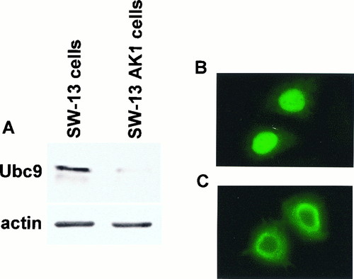
Ubc9 is required for nuclear localization of zVsx-1. (A) Western blot of fractionated cell lysates from SW-13 cells and SW-13 AK1 cells after probing with anti-hsUbc9. SW-13 AK1 cells express A.S.hsUbc9 and show decreased levels of endogenous Ubc9. (B and C) Indirect immunofluorescence using the 9e10 antibody in MT-zVsx-1-transfected SW-13 cells (B) and SW-13 AK1 cells (C). |

Ubc9 restores nuclear localization of zVsx-1 in SW-13 AK1 cells. (A) Indirect immunofluorescence using the 9e10 antibody in SW-13 AK1 cells cotransfected with MT-zVsx-1 and vector. (B) Indirect immunofluorescence using the 9e10 antibody in SW-13 AK1 cells cotransfected with MT-zVsx-1 and Flag-Ubc9. (C) Indirect immunofluorescence with anti-calnexin antibody in SW-13 AK1 cells labels the endoplasmic reticulum. (D) STAT1-NLS-GFP localization in fixed SW-13 AK1 cells. |

Ubc9 C93S also restores zVsx-1 nuclear localization. Indirect immunofluorescence with 9e10 antibody of SW-13 AK1 cells transfected with MT-zVsx-1 and vector control (A), MT-zVsx-1 and Flag-Ubc9 (B), and MT-zVsx-1 and Ubc9C93S (C). |