
|
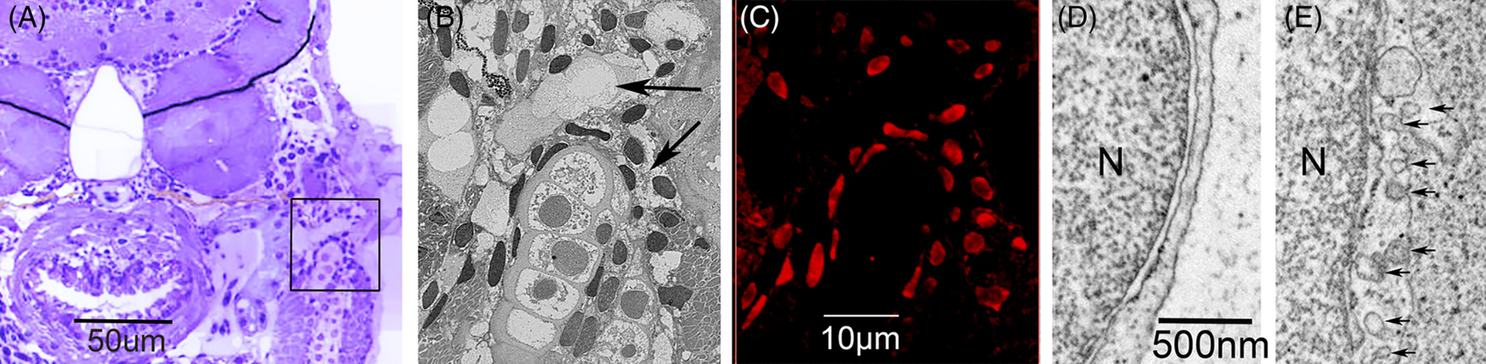
Fig. 7 Cytoplasmic differentiation related to the maturation of the lymphatic endothelium in the zebrafish embryo. At the lymph‐sac of the jugular angle, venous progenitor cells begin to differentiate into functional lymphatic endothelium at 2 dpf and become functional at 9 dpf. A, Cross‐section (toluidine blue) of the trunk to show the lymph‐sac (in black frame) at the jugular angle of the 5 dpf zebrafish. B, Dark‐field SEM image of the lymph‐sac (black arrows) in the frame of Figure 7A. C, Correlative prox1‐immunopositive nuclei of the endothelium lining the lymph‐sac in the frame of Figure 7A. D, TEM images of progenitor cells at 5 dpf. E, The functional lymphatic endothelium at 9 dpf revealed that small pits (or caveola) and coated pits (black arrows) emerged in the cytoplasm of lymph‐progenitor cells after 5 dpf and rapidly increased in number in the functional lymph endothelium. N, nucleus