
Image
|
Figure Caption
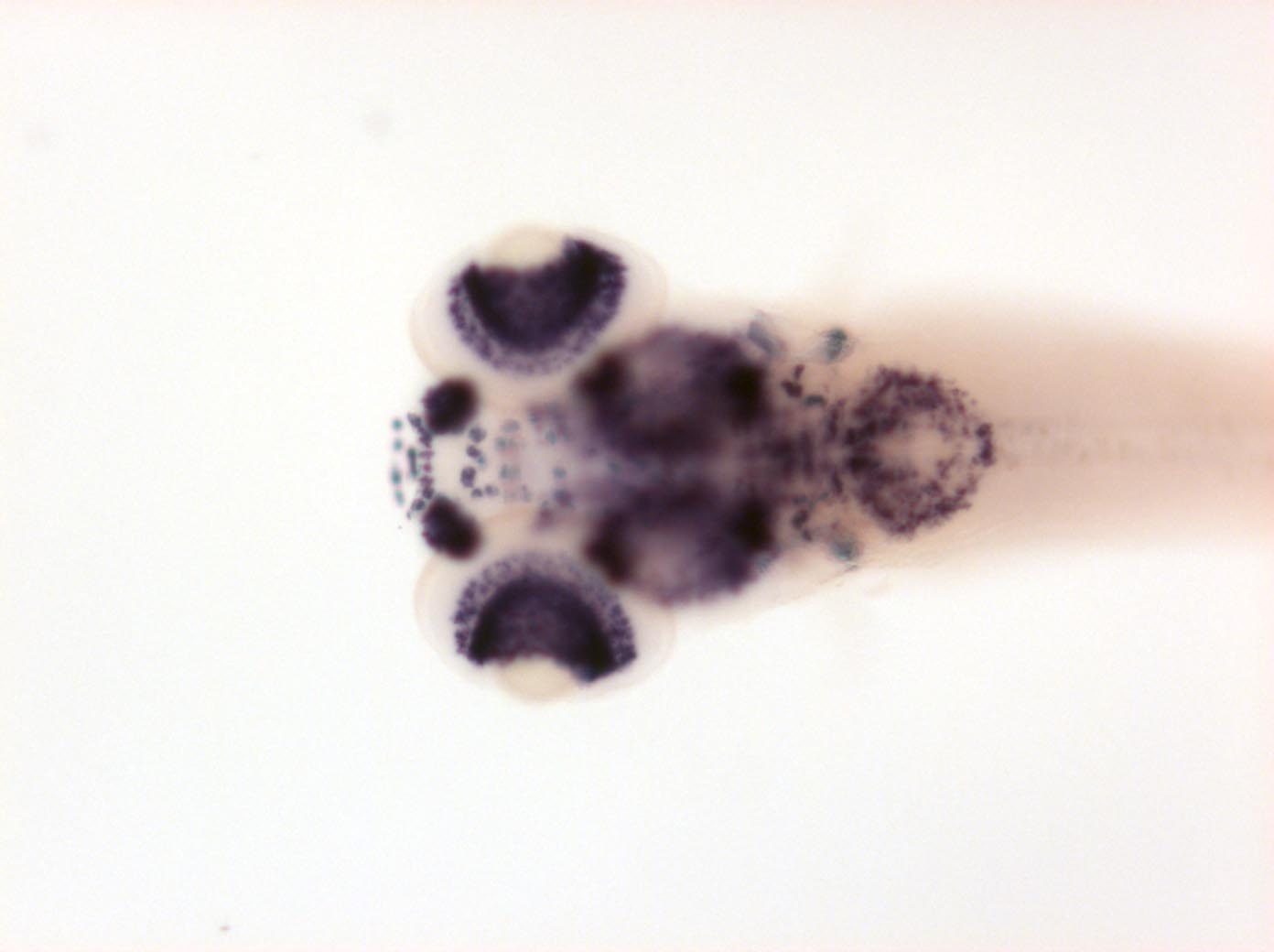
Fig. 5
neurons in olfactory vesicle ++, anterior tectum and tegmentum, cerebellum, ventral lateral hindbrain, spinal cord neurons, ganglion cell layer retina, inner cell layer (weaker), cells in branchial arches, cranial ganglia
Please note that in 5 day old embryos some structures are not accessible to the probe (such as notochord, most of the trunk and tail). Therefore the description of the expression pattern is only partial.
Developmental Stage
Day 5
Orientation
| Preparation | Image Form | View | Direction |
| whole-mount | still | dorsal | anterior to left |
Figure Data