
|
Image description by: Paula Mabee and Nathan Bird
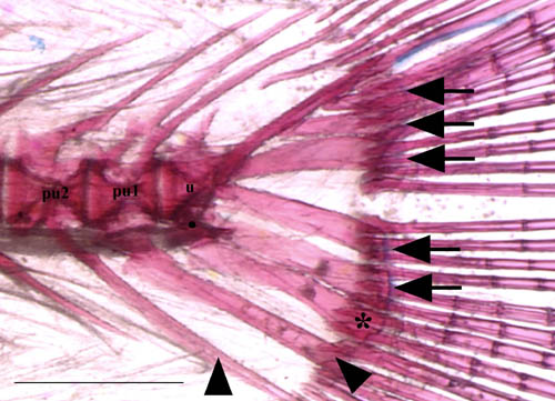
Anatomical structures shown: parhypural, hypurals 1-5, preural centra 1 and 2, urostyle, hemal arches, hemal spines, neural arches, neural spines, parhypurapophysis, lepidotrichia
Stage: adult, 24.4mm SL* *SL (Standard Length) refers to the distance from the tip of the snout (upper or lower jaw, whichever is most anterior) to the posterior edge of the hypurals
Genetic (background) strain: unknown
Genotype: unknown
Animal state: fixed
Labeling: Alcian blue for cartilage, Alizarin red for bone
Description: Image showing skeletal development within the caudal fin of an adult zebrafish, 24.4 mm SL. The parhypural and hypural 1, which form initially as independent condensations (see hypural1.jpeg), fuse proximally. Hypural 2 is continuous with the urostyle. All relevant structures are ossified. Scale bar = 1 mm, arrows = hypurals 1-5, * = parhypural, arrowheads = hemal spines, pu2 = preural centrum 1, pu1 = preural centrum 1, u = urostyle, . = parhypurapophysis
| Preparation | Image Form | View | Direction |
| whole-mount | still | parasagittal | anterior to left |