
|
Image description by: Paula Mabee and Nathan Bird
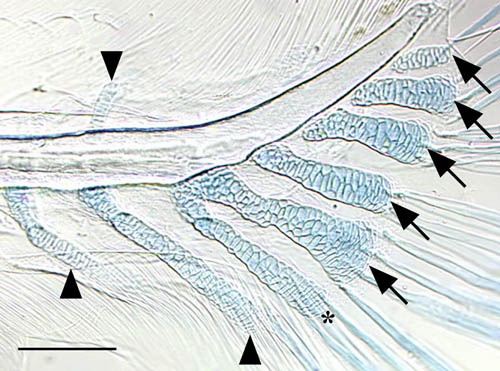
Anatomical structures shown: parhypural, hypurals 1-5, hemal arches and hemal spines of preural centra 1 and 2, neural arches of preural centra 1 and 2, notochord, lepidotrichia, actinotrichia
Stage: larval, 5.8mm SL* (~13 d) *SL (Standard Length) refers to the distance from the tip of the snout (upper or lower jaw, whichever is most anterior) to the posterior edge of the hypurals
Genetic (background) strain: unknown
Genotype: unknown
Animal state: fixed
Labeling: Alcian blue for cartilage, Alizarin red for bone
Description: Image showing skeletal development within the caudal fin of a larval zebrafish, 5.8mm SL. The parhypural and hypural 1, which form initially as independent condensations (see hypural1.jpeg), fuse proximally. Preural centra 1 and 2 and the urostyle are not ossified at this point. Scale bar = 0.1mm, arrows = hypurals 1-5, * = parhypural, arrowheads = hemal spines (ventral to notochord) or neural arch (dorsal to notochord)
| Preparation | Image Form | View | Direction |
| whole-mount | still | parasagittal | anterior to left |