
- Title
-
Fast Homogeneous En Bloc Staining of Large Tissue Samples for Volume Electron Microscopy.
- Authors
- Genoud, C., Titze, B., Graff-Meyer, A., Friedrich, R.W.
- Source
- Full text @ Front. Neuroanat.

Unsuccessful staining attempts. (A) Telencephalon of adult zebrafish stained with a protocol that reduced incubation times of the original BROPA protocol to a total duration of 2 weeks. Note severe tissue damage. (B) Telencephalon of adult zebrafish stained with a protocol that reduced incubation times of the original BROPA protocol to a total duration of 5 days without lead aspartate. Note charging artifacts in nuclei and neuropil. |
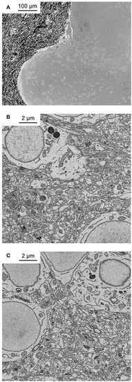
Application of fBROPA to the adult zebrafish brain. (A) Coronal section through the telencephalon of adult zebrafish at the level of Dp (posterior zone of the dorsal telencephalon). Note homogeneous staining. Black particles outside the tissue are silver particles in the surrounding resin to optimize conductivity. (B) Neuropil close to the surface. (C) Neuropil 300 μm below the surface. |

Application of fBROPA to the adult zebrafish brain. (A) Section through the tectum near the location where the diameter is maximal (1.1 mm). Image is a mosaic of 6 × 6 tiles. (B–D) Three images acquired at different depths. Approximate locations of images are indicated by outlines in (A). (E) Examples of images showing synapses (5 nm pixel size). Vesicle pools close to the presynaptic membrane and a thickening of both membranes are visible. Synapse detection can be performed in 3D as shown in Supplementary Data S1 (movie). Note uniformly high contrast. The partial damage on the right side of the tectum occurred during dissection and is unrelated to fixation or staining. |
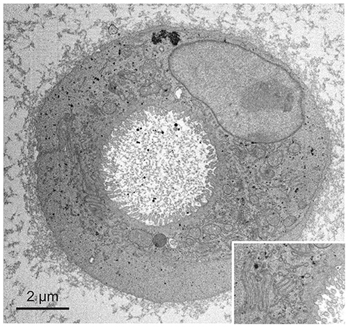
Application of fBROPA to early differentiated organoids (two cells). Insert shows details of the membrane ultrastructure between the two cells. |