
Fig S11
- ID
- ZDB-IMAGE-260821-32
- Publication
- Docampo-Seara et al., 2026 - The microglia-derived protein sema4ab attenuates regenerative neurogenesis after spinal cord injury in zebrafish
- All Figures
- Figures for Docampo-Seara et al., 2026
|
Fig S11
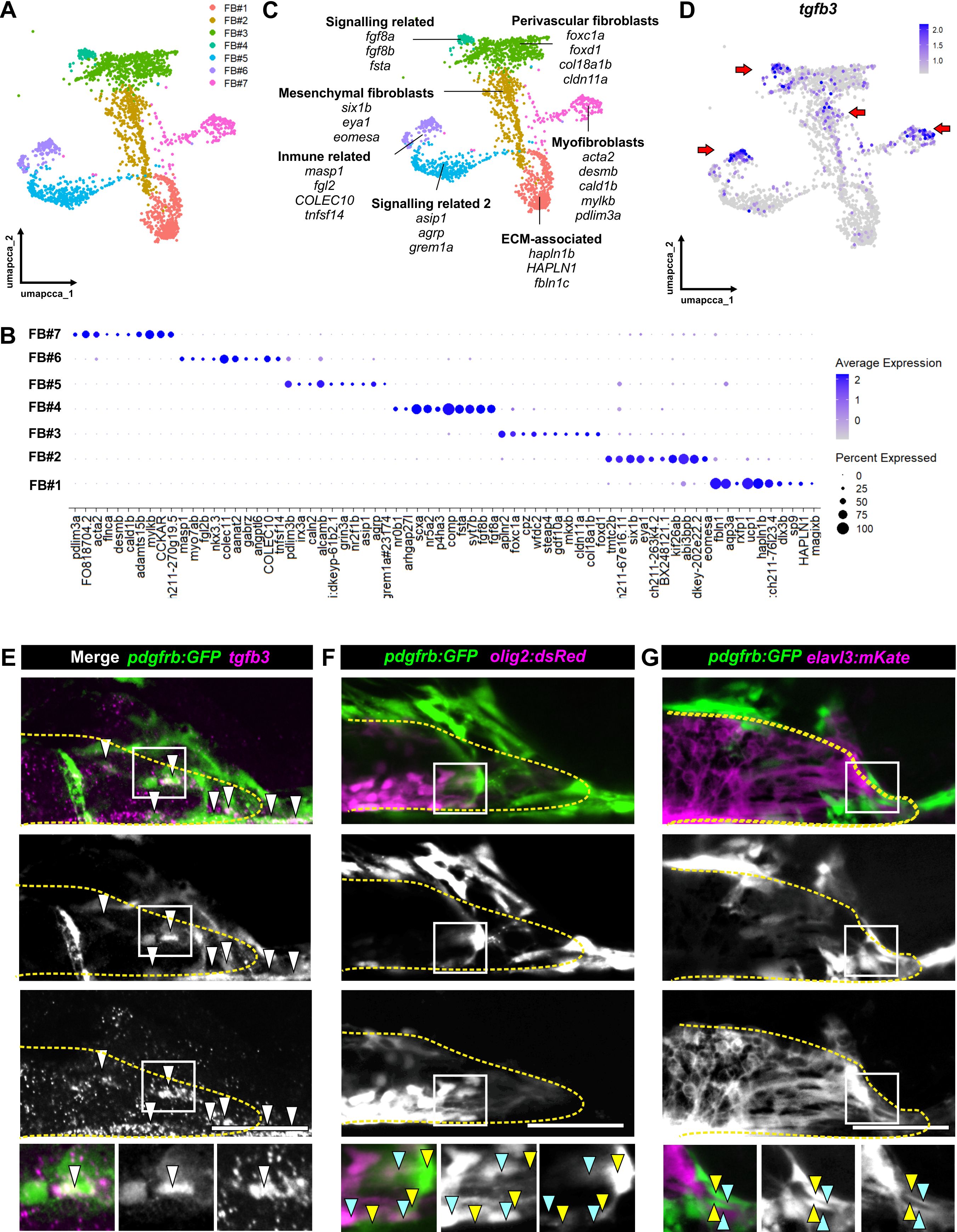
tgfb3 is expressed in subsets of fibroblasts and in close contact with ERGs and growing axons.
(A) UMAP of fibroblasts subset showing 7 clusters of cells. (B) Top 10-ranked gene analysis showing differential expressing genes among clusters. (C) UMAP showing genes used for annotation and tentative cluster names. (D) Feature plot showing expression pattern of tgfb3. Arrows point enriched clusters for the gene. (E) Single-Stacks of the lesion site at the level of the spinal cord showing tgfb3-expressing fibroblasts (arrow). (F) Live imaging showing that fibroblasts (green, yellow arrows) are in the vicinity of motor neuron progenitors (magenta, cyan arrows). (G) Live imaging showing that fibroblasts (green, yellow arrows) are in close contact with growing axons (magenta, cyan arrows). Yellow dotted lines mark the spinal cord. Scale bars: 50 µm.