
Fig S1
- ID
- ZDB-IMAGE-260821-22
- Publication
- Docampo-Seara et al., 2026 - The microglia-derived protein sema4ab attenuates regenerative neurogenesis after spinal cord injury in zebrafish
- All Figures
- Figures for Docampo-Seara et al., 2026
|
Fig S1
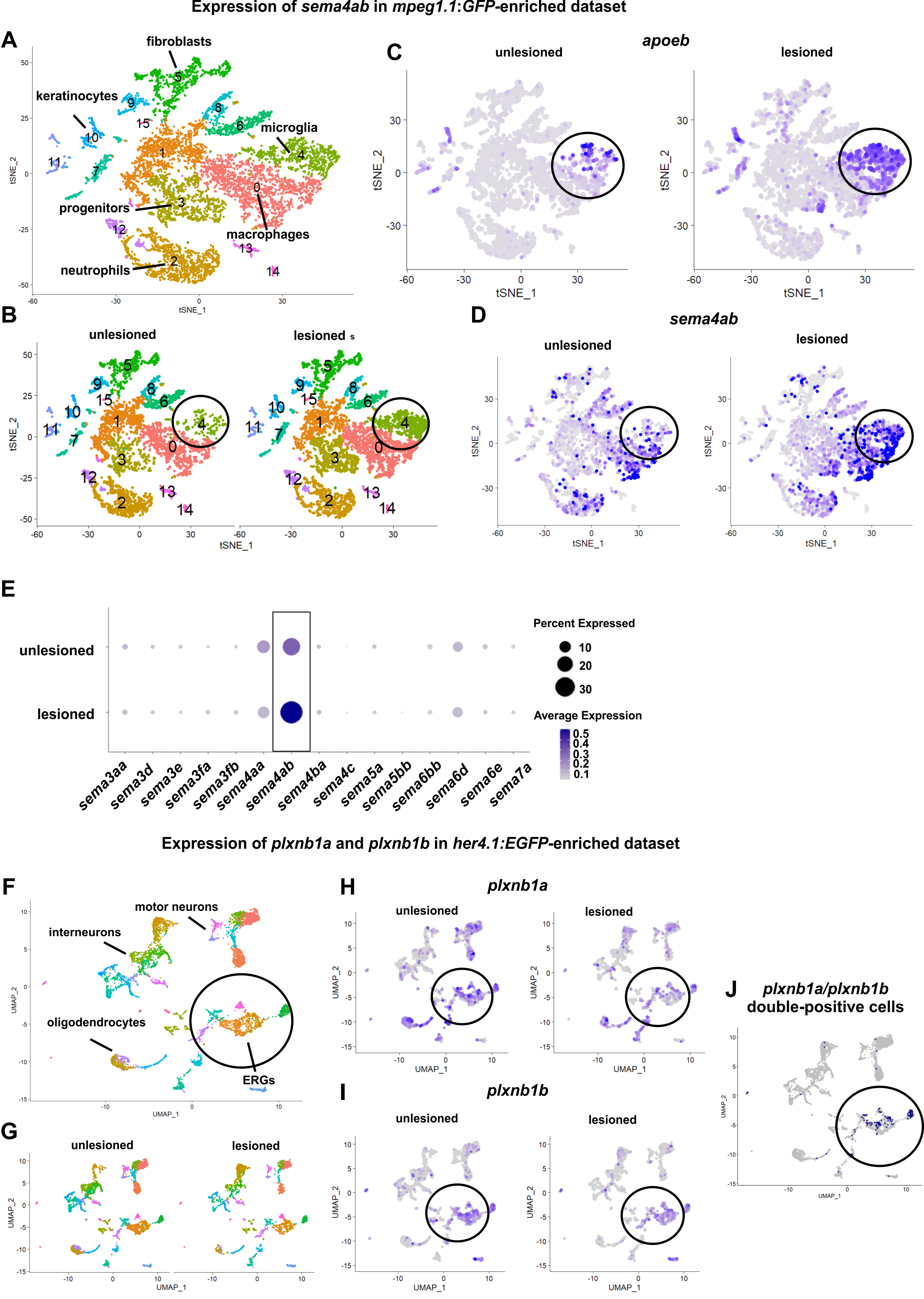
Potential interaction between sema4ab in microglia and plexin receptors in ERGs revealed in cell type-enriched scRNA-seq datasets.
(A) UMAP showing clusters in an mpeg1.1:GFP-enriched scRNA-seq. (B) UMAPs comparing unlesioned and lesioned conditions are shown. Note an increase of the microglia cluster after injury (circle). (C,D) Feature plots comparing expression of apoeb (C) and sema4ab (D) between unlesioned and lesioned larvae, indicating increased presence of both after injury (circled). (E) Dot plot showing all semaphorins expressed in the scRNA-seq dataset before and after lesion. Note that only sema4ab is substantially expressed and also upregulated after injury (rectangle). (F) UMAP showing clusters in a her4.1:EGFP-enriched scRNA-seq. Circle indicates ERGs. (G) UMAPs indicate presence of ERGs in unlesioned and lesioned conditions. (H–J) Feature plots indicating expression of plxnb1a (H) and plxnb1b (I) in ERGs of unlesioned and lesioned larvae (circled). Plotting only cells co-expressing receptors shows strong enrichment in ERGs (J, circled). Data are described in Cavone et al., Dev Cell 2021 Jun 7;56(11):1617–1630.e6.