
Fig. 4
- ID
- ZDB-IMAGE-260805-29
- Publication
- Tian et al., 2026 - Regulation of the PKD2 channel function and associated disease phenotypes by RASSF4
- All Figures
- Figures for Tian et al., 2026
|
Fig. 4
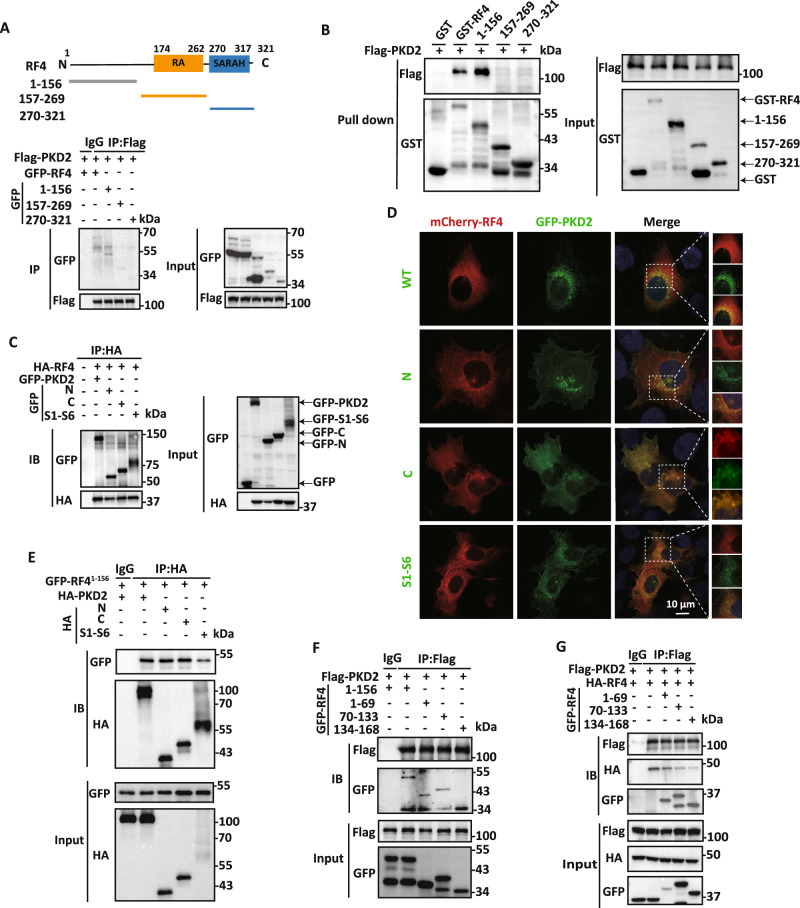
Domains mediating the RASSF4/PKD2 interaction.
A Upper panel, schematic architecture of human RASSF4 protein, with key functional domains indicated. Lower panels, representative co-IP data from HEK cells, showing interaction of Flag-PKD2 with GFP-RASSF4 and three GFP-tagged RASSF4 fragments. B Representative data obtained from GST pull-down assays showing the RASSF4/PKD2 interaction. HEK cells expressing Flag-PKD2 were incubated with purified GST-RASSF4 truncation proteins. Complexes were pulled down with glutathione beads and analyzed by WB. C Representative data from co-IP assays in HEK cells co-expressing HA-RASSF4 with different GFP-tagged PKD2 fragments (left panels) or co-expressing GFP-RASSF4 with different HA-tagged PKD2 fragments (right panels). “N”, PKD2 N-terminus (M1-S219), “C”, C-terminus (I671-V968), and “S1–S6”, central domain (K215-M675). D Representative confocal microscopy images showing subcellular colocalization in U-2 OS cells of mCherry-RASSF4 with different GFP-PKD2 constructs, as indicated. Scale bar: 10 μm. E Representative co-IP data showing interaction of GFP-tagged RASSF4-M1-P156 with different HA-PKD2 fragments, as indicated, in HEK cells. F, G Representative co-IP data showing interaction of Flag-PKD2 with different GFP-tagged RASSF4 fragments, as indicated, in HEK cells.