
Fig. 1
- ID
- ZDB-IMAGE-260805-26
- Publication
- Tian et al., 2026 - Regulation of the PKD2 channel function and associated disease phenotypes by RASSF4
- All Figures
- Figures for Tian et al., 2026
|
Fig. 1
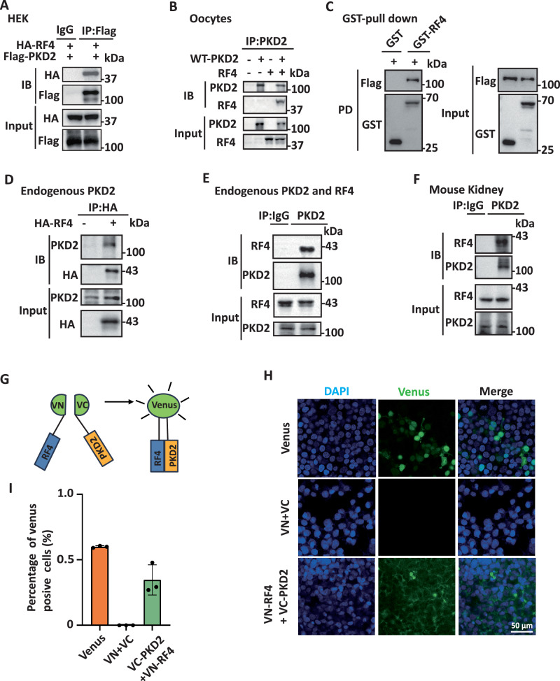
Physical interaction between PKD2 and RASSF4.
Representative data showing interaction between exogenous RASSF4 (RF4) and PKD2 in HEK cells (A) and Xenopus oocytes (B) detected using co-IP assays and WB. C Representative data showing interaction between purified GST-tagged RASSF4, expressed in E. coli BL21, and Flag-tagged PKD2, expressed in HEK293T cells, assessed by GST pull-down assays. Representative data showing interaction of over-expressed (D) and endogenous (E) RASSF4 with endogenous PKD2 in HEK cells, examined using co-IP. F Representative data showing interaction between endogenous RASSF4 and PKD2 assessed by co-IP in a kidney of an 8-week-old C57BL/6 mouse. G–I The RASSF4/PKD2 interaction was analyzed using BiFC assays. G Schematic presentation of a BiFC assay. The Venus N-terminal half (VN) and C-terminal half (VC) were tagged to the N-terminal end of RASSF4 and PKD2, respectively, for transfection in HEK cells. The green fluorescent signal indicates proximity between VN and VC, which indicates the presence of the RASSF4/PKD2 association. H Representative data showing fluorescence in HEK cells transfected with Venus, VN + VC or VN-RF4 + VC-PKD2. Nuclei were stained with DAPI. I Statistical data showing quantified fluorescence intensities obtained under the same conditions as in (H).