
FIGURE 2
- ID
- ZDB-IMAGE-260626-15
- Publication
- Rezi et al., 2026 - ZDHHC5 interacts physically and functionally with DLG1 at primary cilia and regulates ciliary length and kidney morphology
- All Figures
- Figures for Rezi et al., 2026
|
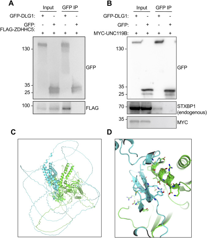
FIGURE 2
DLG1 binds to ZDHHC5 and STXBP1. (A,B) HEK293T cells co-expressing GFP-DLG1 or GFP alone (negative control) with FLAG-ZDHHC5 (A) or MYC-UNC119B (B) were subjected to IP with GFP beads, and input and IP pellets analysed by western blotting using antibodies as indicated. Molecular mass markers are shown in kDa to the left. (C,D) Structural model of the DLG1-ZDHHC5 interaction. (C) AlphaFold2-Multimer model showing the predicted structure of the complex between DLG1 (green) and ZDHHC5 shown in cartoon representation. In addition to the structured domains, large regions are predicted to be disordered. (D) Zoom-in on the interface between the GUK-like domain (residues 715–890) of DLG1 and ZDHHC5’s N-terminal region (residues 70–250). Interface residues with PAE < 4Å are highlighted as sticks.