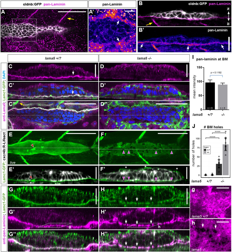
Laminin α5 is an integral component of the pLLP migration substrate. (A-B′) Pan-laminin immunostaining in whole-mount embryos (A,A′) and transverse cryosections (B,B′). Yellow and white arrows point to somite boundaries and the epidermal BM, respectively. Additional magnifications from A are in Fig. S8A-A″. (C-D″) Lama5 immunostaining in control (lama5+/?) (C-C″) and lama5−/− (D-D″) lamc1-GFP embryos. Orthogonal views (C-D′) and single z-planes (C″,D″) show Lama5 and Lamc1 in the epidermal BM (white arrows in C,C′) in controls. Lama5, but not Lamc1, is missing in the BM in lama5 mutants (D-D″). (E-F′) Live imaging of lamc1-GFP highlighting BM holes in lama5 mutants (purple arrowheads). (E-F′) Maximum-intensity projections of 5 μm substacks (E,F) and single orthogonal views (E′,F′). Lamc1-GFP also labels the Schwann cells of the PLLn (red arrowheads). (J) Quantification of the number of holes underneath pLLP (dark gray) and adjacent skin (light gray). (G-H″,g,h) Pan-laminin immunostaining in lamc1-GFP embryos. Orthogonal views (G-H″) and higher magnifications of single z-planes (g,h, as indicated in G′,H′), show similar discontinuities in the BM in lama5 mutants (white arrows in H-H″, h). (I) Quantification of pan-laminin signal intensity (data are mean±s.d.). (J) Dots represent data from individual embryo (data are mean±s.d.) Unpaired, two-tailed t-test; ****P<0.0001. Scale bars: 50 μm for top-views in A,C″,D″,E,F; 20 μm for optical sections in B,B′,C,C′,D,D′,E′,F′,G-H″; 10 μm for higher magnifications (A′,g,h). a, apical; b, basal.
Acknowledgments
This image is the copyrighted work of the attributed author or publisher, and
ZFIN has permission only to display this image to its users.
Additional permissions should be obtained from the applicable author or publisher of the image.
Full text @ Development
Your Input Welcome
Thank you for submitting comments. Your input has been emailed to ZFIN curators who may contact you if
additional information is required.
Oops. Something went wrong. Please try again later.