
Fig. 3
- ID
- ZDB-IMAGE-260617-9
- Genes
- Publication
- Martin et al., 2026 - Nr2f1a and Isl1 repress acquisition of epicardial identity in venous atrial cardiomyocytes
- All Figures
- Figures for Martin et al., 2026
|
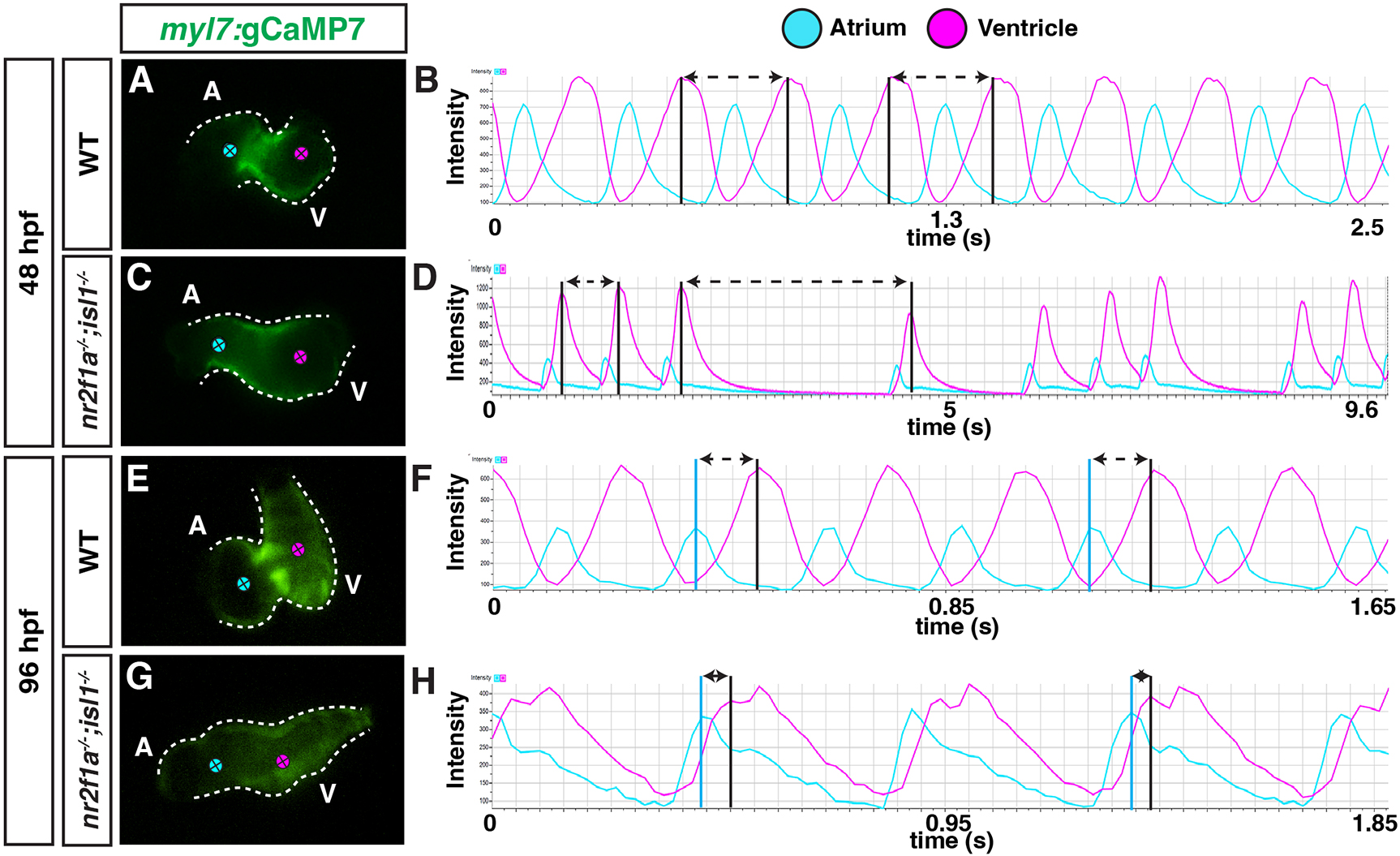
Fig. 3 nr2f1a;isl1 mutants functionally lack pacemaker cardiomyocytes. (A,C,E,G) Images from videos of wild-type and nr2f1a;isl1 mutant hearts carrying the myl7:jGCaMP7c transgene at 48 and 96 hpf. Magenta and blue dots indicate the location of jGCaMP7c fluorescence recordings in the ventricles and the atria, respectively. (B,D,F,H). Intensity traces of myl7:jGCaMP7c fluorescence from the ventricles (magenta) and atria (blue). Arrows between black lines in B and D indicate the time between maximum intensity of calcium waves in the ventricle. Arrows between blue and black lines in F and H indicate the time between maximum atrial intensity and initial peak in the ventricle. 48 hpf: wild type (n=5) and nr2f1a−/−;isl1−/− (n=3); 96 hpf: wild type (n=5) and nr2f1a−/−;isl1−/− (n=3).