
|
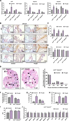
Fig. 7 Phenotypic, biochemical, and molecular parameters in WT (amh+/+;bmpr2a+/+), amh±;bmpr2a+/+, amh+/+;bmpr2a±, amh±;bmpr2a± female zebrafish after 150-d exposure to neburon. (A) The changes in transcriptional levels of amh, bmpr2a, and bmpr2b (n = 4 samples/group). (B) Representative ovarian sections were labeled with pSmad1 using immunostaining. Areas of positive signal are magnified in the right panel. Scale bars represent 200 μm (left panel) and 40 μm (right panel). (C) The proportion of pSmad1-positive (pSmad1+) area to total follicles area in ovarian sections (n = 4 samples/group). (D) The representative ovary section stained with H&E. The letters PO, CO, VO, MO, and AF next to the triangle, arrow, star, dagger maker, and pound sign indicated primary oocyte, cortical alveolar oocyte, vitellogenic oocyte, mature oocyte, and atretic follicle, respectively. Scale bar = 500 μm. The proportion of the number of oocytes from each stage in each group after neburon treatment (n = 4 samples/group). (E) The AMH levels in the muscle of female zebrafish after 150-d exposure to neburon (n = 5∼6 samples/group). Average total number of spawned embryos (n = 10 females/group) (F), malformation rate of F1 larvae (n = 6 dishes/group) (G), and mortality of F1 larvae (n = 6 dishes/group) (H). (I) Gonadosomatic index (GSI) in amh±;bmpr2a± female zebrafish (n = 10 samples/group) exposed to neburon for 150 d. (J) The mRNA expression of igfs-related genes (n = 4 samples/group). Data are presented as the mean ± SE. Statistical analysis was analyzed by t-test between two groups. *p < 0.05, **p < 0.01, and ***p < 0.001. The data presented in Figure 7A and 7C-J are also displayed in Excel Table S27 and S28–35, respectively.