
Figure 8
- ID
- ZDB-IMAGE-260508-27
- Publication
- Pannese et al., 2026 - Impaired collagen deposition in the absence of ERp44
- All Figures
- Figures for Pannese et al., 2026
|
Figure 8
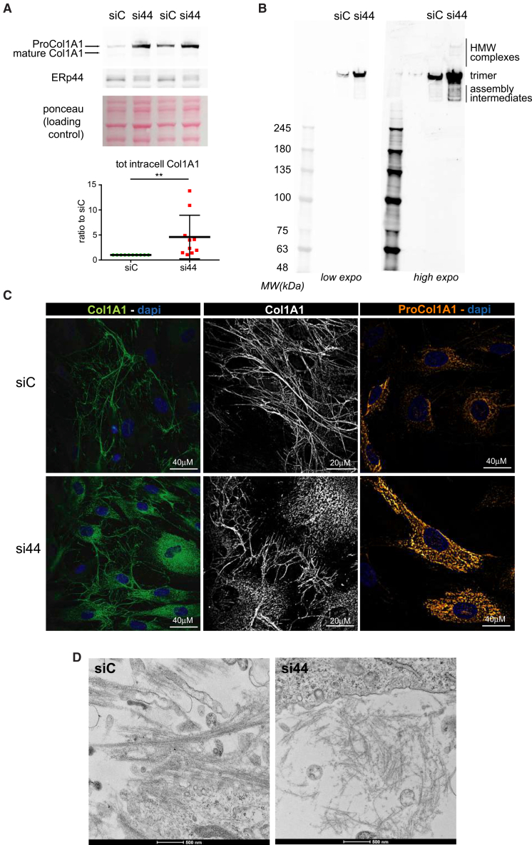
ERp44 KD induces intracellular accumulation of ProCol1A1, as well as defective extracellular collagen deposition in human fibroblasts (BJ cells)
BJ cells were subjected to silencing of ERp44, or treated with control duplexes (si44 and siC, respectively). About 72 h after silencing, protein extracts were collected.
(A) Representative immunoblot images of Col1A1 and ERp44 proteins with the corresponding Ponceau-stained membrane; samples were separated on a 4%–12% pre-cast SDS-PAGE gel under reducing conditions in MOPS (3-(N-morpholino)propanesulfonic acid) buffer. Membrane were probed with anti-Col1A1 antibody (
(B) Protein extracts of ERp44 silenced (si44) and control (siC) BJ cells were separated on a 4%–12% precast SDS-PAGE gel under non-reducing conditions in MOPS buffer. Membranes were acquired at two different laser intensity (low and high exposition). Under high exposition, si44 sample (mean of ERp44 downregulation), reveals the accumulation of collagen high molecular weight (HMW) complexes, as well as collagen covalent assembly intermediates.
(C) Immunofluorescence images of siC (top) or si44 (bottom) BJ cells. Left: cells were fixed in methanol and then stained, without additional permeabilization, with anti-Col1A1 antibody (
(D) Transmission electron microscopy images of siC (left) or si44 (right). BJ extracellular collagen fibers: long and straight collagen fibers are deposited extracellularly in siC cells in contrast to thinner and disorganized collagen fibers in ERp44 KD cells (si44). Scale bar 500 nm.