
Figure 3
- ID
- ZDB-IMAGE-260508-22
- Publication
- Pannese et al., 2026 - Impaired collagen deposition in the absence of ERp44
- All Figures
- Figures for Pannese et al., 2026
|
Figure 3
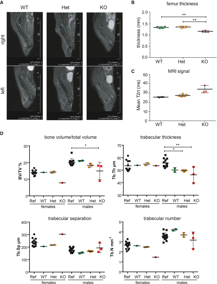
Femoral long bone mineralization delay in ERp44 KO mice
(A) MRI images of right (3A, top) and left (3A, bottom) femurs of WT, Het, and KO ERp44 male littermates of 11 weeks of age.
(B) Bone thickness quantification of left and right femurs siblings at 11 weeks of age (
(C) MRI T2rt signal is higher in KO mice (
(D) μCT analysis of femurs from siblings at 11 weeks of age and reference mice of the same age and sex: decrement of bone volume fraction, trabecular thickness, and trabecular number, slight increment of trabecular separation in KO animals. Average ± SD, One-way ANOVA and Bonferroni’s multiple comparison test (∗