
Figure 2
- ID
- ZDB-IMAGE-260508-19
- Publication
- Pannese et al., 2026 - Impaired collagen deposition in the absence of ERp44
- All Figures
- Figures for Pannese et al., 2026
|
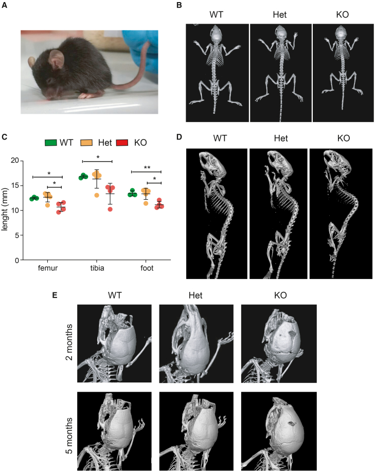
Figure 2
ERp44 KO mice display skeletal anomalies by means of CT scan
(A) Representative phenotype of ERp44 KO mouse at 2 months of age; note the exacerbated column curvature.
(B) Skeleton top view of three littermate mice (females) at 2 months of age.
(C) Femoral, tibial, and foot length quantification of the three genotype mice at 11 weeks of age (
(D) Skeleton lateral view of the same mice in
(E) Skull of 2 month-old mice (females): note open cranial suture of the frontal bones in KO mice (2E, top), which remains partially open at 5 months of age (2E, bottom).